
How Technology Is Changing How We Treat Bulk Billing Imaging
Types Of Tests
Content
- Imaging And Also Radiology Services We Offer:.
- What To Expect.
- New Medicare Guidelines For Innovative Imaging Requests Will Certainly Call For Considerable Resources.
- Qualifying Solutions.
- Upcoming Services.
Imaging And Radiology Solutions We Offer:.
Please fill in the on the internet request form on the EMR Interface Request Kind Web page or call Caitlin Chin at. No, this will certainly lead to the wrong test being arranged as well as potentially performed. Cigarette smokers between the ages of with a tobacco-smoking history are urged to get regular testing for lung cancer.
Service providers are motivated to review all NCDs and also LCDs for services provided to guarantee all documentation demands are met. Most commercial payers require that analysis tests such as CT, MRI, and PET DOG be precertified. It is the referring doctor's obligation to get this precertification by contacting the payer and also offering the clinical factor for the test, a function generally performed by their nursing staff. Upon authorization, the payer will provide a precertification/preauthorization number, which need to be submitted by both the center as well as the translating medical professional. If the payer refuses to approve the test, neither the facility neither the doctor will be paid for it regardless of the findings. In some cases it is much easier to review what you can do as opposed to what you can not.
- Have previous studies readily available, preferably, to try to find adjustments vs. standard.
- Many CT checks in cancer people are to assess the chest, hips, and abdominal area.
- They are utilized for detecting and staging, assessing treatment feedback, and also checking for reoccurrence.
- At JADPRO Live at APSHO, Dr. Steele explained the ABCs of diagnostic radiology, describing which tests are best for which situations, as well as which are a misuse of health care bucks.
An evaluation of the blood flow to the wall surfaces of your heart making use of a high resolution ANIMAL scanner. A treatment in which the blood supply of uterine fibroids is cut off to obtain them to diminish.
What To Anticipate.
" This is reaching be a trouble, as our treatments are improving. Our people are currently living past three PET/CTs," Dr. Steele commented. Cancer clients usually have refined searchings for and chest x-rays may be also fundamental to select them up, he discussed. " Breast x-rays as well as mammography have very little radiation," he claimed. " With any luck, with this information you can lessen your person's worries." Having actually a set up test in non-emergency scenarios is very important, because lots of imaging tests require unique individual prep.
This procedure increases performance as well as precision which lowers clinical mistakes as well as provides safer care for the individual. Like many other types of diagnostic imaging, MRIs are a painless, noninvasive means for a physician to see plainly into your back as well as find the source of your discomfort. Some patients stress they will certainly really feel claustrophobic or nervous during an MRI. At Envision Imaging, we make use of the most up to date MRI innovation, which is not as restricting as outdated tools. Additionally, our engineers are trained to help you unwind as well as really feel as comfy as feasible during the procedure. At McLaren https://brightonradiology.com.au/imaging-services/ct-guided-injections/ we utilize the current imaging equipment for our patient CT scans.
I guaranteed those at the center that the recognized concerns might be fixed, yet that it would certainly be a strenuous process to obtain settlement on appeal for those solutions that had already been denied. Failing to do so could result in loss of compensation for both the facility as well as the physician. For industrial payers that do not require precertification, there may be some versatility in the transforming of orders; however, it is tough to track this details on a department level. This is a primary reason most facilities have picked to execute a conventional plan that treats all payers the same and also does not call for the technologist to review economic information.
New Medicare Rules For Sophisticated Imaging Requests Will Certainly Need Substantial Sources.
Make use of our https://en.search.wordpress.com/?src=organic&q=radiology open MRI systems for a comfortable, hassle-free experience. You'll value open MRI equipment that does not enclose you throughout the check. Calm, spacious MRI areas allow enough for a parent or assistance person. Rely upon our team of board-certified radiologists to examine your examinations and also identify your condition. Your experienced radiologist will supply your imaging results to your referring physician and also speak with on your therapy if required. If you get an x-ray for pain in the back at an Envision Imaging center, you can expect a particularly trained engineer to execute the treatment as successfully as feasible as well as ensure you fit throughout. Pain in the back is a common reason people miss work or see their medical professional.
Use of a rubber stamp trademark is not an approved method of authentication. Checking facilities ought to not consistently modify analysis examination orders from the buying medical professional.
Upcoming Solutions.

An x-ray mostly aids doctors identify bone problems and is a faster treatment than an MRI. New government regulations will certainly dictate when as well as exactly how medical professionals can buy advanced imaging tests for Medicare people to avoid unnecessary treatments and suppress medical care spending. Your doctor might get an imaging test to identify the factor for your back pain. Tests such as x-rays, CT scans and MRIs allow a doctor to check out your back without executing surgery.

20 Reasons You Need To Stop Stressing About Is A Cat Scan An X Ray
Diagnostic Radiology
Content
- Imaging And Radiology Solutions We Supply:.
- What To Expect.
- Brand-new Medicare Rules For Sophisticated Imaging Demands Will Need Considerable Sources.
- Qualifying Solutions.
- Upcoming Solutions.
Imaging And Radiology Services We Provide:.
Lab researches of blood, urine, or joint fluids are utilized to identify the presence and also amount of chemicals, proteins, as well as other compounds. Your physician may get different laboratory studies depending on what she or he discovers during the preliminary exam.
The Centers for Medicare & Medicaid Providers stipulates separate demands for diagnostic test orders, relying on where the testing takes place. People need to complete this release to allow Augusta Health and wellness to request past imaging from other facilities where they have had imaging done. If this is your very first time referring an individual to Stanford for imaging, please full theUnknown Supplier Form for the Medical Personnel Workplace supplier database. The reading room aide is readily available to link you with our sub-specialty radiologists and technologists. Call us to set up an appointment at any one of our imaging places.
- As long as the https://brightonradiology.com.au/imaging-services/ct-guided-injections/ hospital regulating body has accredited radiologists to buy services for patients, then the order stands under the POLICE.
- Bust MRI scans ought to be arranged within 7-12 days of the onset of one's menstrual cycle unless the demand is immediate.
- The MRI maker makes use of a large magnet and also a computer to take images of the within your body.
- If an order is transformed from what was initially requested, the clinical necessity of the adjustment likewise must be recorded to support the modified order.
- As with nonhospital imaging, orders need to consist of the patient's name, the examination asked for, scientific signs for the test, as well as the name and trademark of the treating doctor.
Ultrasonography, which is often called sonography, uses high-frequency sound waves and also a computer system to develop pictures. The main use of breast ultrasound today is to help diagnose bust abnormalities detected throughout a physical examination as well as to define prospective abnormalities seen on mammography or breast. Say "no thanks" to exorbitant health center imaging rates and charges.
What To Anticipate.
" This is getting to be a problem, as our therapies are getting better. Our clients are currently living past 3 PET/CTs," Dr. Steele commented. Cancer patients usually have refined findings and also upper body x-rays may be as well rudimentary to pick them up, he described. " Upper body x-rays and also mammography have extremely little radiation," he stated. " Ideally, with this details you can allay your client's fears." Having an arranged test in non-emergency situations is essential, considering that many imaging tests call for special person prep.
Magnetic vibration imaging is utilized to aid direct the radiologist's instruments to the website of the uncommon growth. You can message your center, sight lab results, schedule a consultation, as well as pay your costs. Work together with radiologists and also referring medical professionals to improve imaging appropriateness based on Picking Carefully topics.
CMS has actually specified that a trademark is not called for on orders for tests paid under the professional lab or medical professional cost routine. The radiologist might cancel an order when the problem of the person avoids conclusion of the bought diagnostic examination. There are a few circumstances in which the testing center may supply various or added tests. Perhaps the most crucial point in the above 5 criteria is the one stating that postponing performance of the test would have a damaging result on the person.
New Medicare Guidelines For Sophisticated Imaging Demands Will Require Considerable Resources.
It is important to bear in mind that the Centers for Medicare & Medicaid Providers guidelines for independent analysis testing facilities/physician workplaces differ from hospital guidelines. Nevertheless, several exclusive payers do not distinguish their standards based on the location of service. Private payer demands for orders differ considerably, and it is very important to integrate an approach for resolving these variations.
Certifying Solutions.
Use a stamp signature is not an authorized approach of authentication. Checking centers must not regularly customize analysis test orders from the purchasing medical professional.
As a result, it is important that your management reviews that is accountable for making certain correct orders and that you do not wrongly overburden your medical staff with this heavily administrative responsibility. There is terrific value in having clinical team evaluation the orders, but that does not convert into giving them the main obligation for getting right orders. While we would really like this to be a precise problem, it is not.

20 Insightful Quotes About Doctors That Do X Rays Near Me
Types Of Examinations
Content
- Imaging As Well As Radiology Solutions We Supply:.
- What To Expect.
- New Medicare Policies For Innovative Imaging Requests Will Call For Substantial Sources.
Imaging https://brightonradiology.com.au/imaging-services/ct-guided-injections/ And Radiology Solutions We Supply:.
MRIs are excellent for identifying soft cells and also spine tendon issues. Superior imaging technology as well as specialized medical professional testimonial establishes a structure for concentrated treatment.
Service providers are urged to check out all NCDs as well as LCDs for services supplied to make certain all documentation demands are fulfilled. Many business payers require that diagnostic tests such as CT, MRI, as well as PET be precertified. It is the referring physician's obligation to get this precertification by getting in touch with the payer and supplying the clinical factor for the test, a feature generally performed by their nursing personnel. Upon approval, the payer will issue a precertification/preauthorization number, which have to be submitted by both the facility as well as the translating doctor. If the payer rejects to authorize the examination, neither the center nor the medical professional will certainly be paid for it no matter the findings. Often it is less complicated http://edition.cnn.com/search/?text=radiology to review what you can do as opposed to what you can't.
- As long as the health center controling body has licensed radiologists to purchase solutions for clients, after that the order stands under the POLICE OFFICER.
- Magnetic resonance imaging is a noninvasive medical checkup that does not use ionizing radiation (X-rays).
- Bust MRI scans need to be scheduled within 7-12 days of the start of one's menstrual cycle unless the request is urgent.
- The MRI maker utilizes a huge magnet and a computer to take pictures of the within your body.
- If an order is altered from what was originally requested, the clinical requirement of the change additionally ought to be documented to sustain the revised order.
- As with nonhospital imaging, orders must consist of the individual's name, the examination requested, scientific indicators for the test, and also the name as well as trademark of the dealing with doctor.
The analyzing medical professional plainly papers why added examinations were executed. The result is communicated to dealing with doctor and made use of in therapy. Regardless of setup, remember that the getting physician is in charge of recording medical necessity for a test order. The guidelines regulating IDTFs are the most details and also stringent due to abusive billing practices that were running rampant many years earlier, when IDTFs were notorious for routinely including tests that were not bought or not medically essential. If the referring physician shows a "rule out," the indicators or signs motivating the test that ruled out the problem must be consisted of in the paperwork.
What To Anticipate.
" This is reaching be an issue, as our therapies are getting better. Our patients are currently living beyond 3 PET/CTs," Dr. Steele commented. Cancer cells people often have subtle findings and breast x-rays may be too fundamental to pick them up, he clarified. " Breast x-rays as well as mammography have very little radiation," he stated. " Hopefully, with this details you can abate your person's anxieties." Having a scheduled test in non-emergency situations is necessary, given that numerous imaging examinations call for special person prep.
Virginia Mason is dedicated to maintaining a secure setting for clients, their families and also site visitors. Oregon Health & Science College is dedicated to enhancing the health and also lifestyle for all Oregonians through quality, advancement and also management in health care, education and learning as well as study. Outdoors medical professionals as well as centers can buy our tests with this downloadable type. We offer a vast spectrum of diagnostic and also therapeutic solutions in a reliable, professional and also prompt fashion.
At OhioHealth, our imaging as well as radiology specialists utilize innovative innovations to aid identify and deal with various conditions. Every one of our radiologists have accessibility to past reports from all OhioHealth imaging places, making accurate record comparisons easy, regardless of which location you select. You will certainly require a manuscript from your physician, except for routine testing mammography. Your medical professional will provide the prep instructions you require for your imaging examination, so you feel ready for your appointment. If your test consists of an IV shot of a comparison color, you will be instructed not to consume or drink anything for three hours prior to your test.
Brand-new Medicare Policies For Innovative Imaging Requests Will Call For Substantial Resources.
Make use of our open MRI devices for a comfortable, hassle-free experience. You'll appreciate open MRI tools that doesn't confine you throughout the scan. Calm, large MRI areas are big sufficient for a parent or assistance person. Rely on our team of board-certified radiologists to review your tests as well as identify your condition. Your experienced radiologist will provide your imaging outcomes to your referring doctor and speak with on your treatment if needed. If you get an x-ray for pain in the back at an Envision Imaging center, you can expect a specifically trained technologist to perform the treatment as efficiently as possible and ensure you are comfortable throughout. Neck and back pain is a typical factor individuals miss job or see their doctor.
Despite the confirming physician, a modified order should be acquired to make sure that the research executed matches the study ordered. AnMRI and also x-rayare both pain-free treatments and also useful analysis tools, and every one has advantages. As an example, an MRI is a much better tool for identifying soft cells troubles, as well as it does not subject clients to ionizing radiation.
Upcoming Solutions.
Clinical tests are study studies that review a new clinical method, gadget, medication, or various other therapy. As a Stanford Health Care patient, you might have accessibility to the most up to date, advanced clinical tests. Tomosynthesis utilizes reduced dose x-rays to take mammogram images of the breast, as well as shows only a few layers of the breast at a time. Preliminary researches show higher cancer discovery and also reduced incorrect positives than full-field electronic mammography. A details sort of imaging that uses a low-dose x-ray system to analyze breasts and also help in the very early discovery and medical diagnosis of bust illness in females.

The Evolution Of Bulk Billing X Ray Near Me
Radiology As Well As Pathology Solutions
Content
- Imaging And Radiology Services We Give:.
- What To Expect.
- Brand-new Medicare Rules For Advanced Imaging Requests Will Certainly Call For Significant Resources.
Imaging And Radiology Services We Provide:.
While this write-up will certainly concentrate on the CMS as well as private payers in a broad feeling, practices should bill per specific payer standards. The CMS has actually released details regulations in the Medicare Advantage Policy Handbook (phase 15, section 80.6) for the buying of analysis examinations (/ manuals/Downloads/bp102c15. pdf).
Your doctor has asked for a computed tomography check of your abdomen and hips. CT scans usage X-ray technology and also progressed computer system evaluation to produce in-depth photos of your body.

- X-rays are usually the first type of examination a physician will order to dismiss bone injuries.
- It has been estimated that $12 billion is https://brightonradiology.com.au/imaging-services/ct-guided-injections/ wasted yearly on unnecessary imaging.
- Obtaining a back x-ray is a fast, pain-free and also noninvasive treatment.
The C-View software program alternative creates synthesized 2D images from tomosynthesis information collections. C-View pictures may be used with tomosynthesis in the screening and also medical diagnosis of bust cancer cells, removing the need for a separate 2D direct exposure. The radiation dosage with tomosynthesis and also C-View uses the medical benefits of tomosynthesis at concerning the same ordinary dose of 2D electronic mammograms in the U.S.A.. In order to inform individuals, it's great to understand the approximate amount of ionizing radiation the various tests discharge. " There's whole lots in journalism regarding this, as well as lots of misunderstanding and also person fear," he claimed. Dr. Hentel and coworkers state that durable proof supporting the wanted outcome of decreasing unacceptable sophisticated imaging is lacking until now.
What To Anticipate.
Trust our knowledgeable imaging group and also board-certified radiologists at Wayne UNC Healthcare to create premium pictures and also supply exact outcomes. You'll have access to advanced modern technology as well as practical services close to house in Goldsboro, North Carolina. Your medical professional could ordera CT scanto diagnose a back injury pertaining to soft tissues. A CT check programs pictures of muscular tissues, organs and ligaments more clearly than a traditional x-ray. CT scans are additionally typically utilized in action to an emergency such as a crash or other injury. Neck and back pain is hardly ever an indication of a serious underlying problem, however it can be.
A CT check of the abdominal area as well as hips can aid identify issues in the bladder, womb, prostate, liver or bowels. At Envision Imaging, we recognize just how difficult it is to handle neck and back pain, and also we desire you to know you're not alone. Our knowledgeable as well as caring group is ready to aid you locate the source of your pain, so you can start the right treatment and also proceed with your life. Making use of first-rate analysis tools, we intend to create a pleasant, practical as well as affordable experience for our patients each time without sacrificing high quality or accuracy. To read more concerning our imaging solutions, connect to our team orschedule a visit today. CT scans are the favored tool for diagnosing extreme injuries that need prompt attention, and also they are likewise useful in locating tumors. Nevertheless, they expose patients to a little dosage of radiation, whereas MRIs do not.
Monitoring needs to make sure that the ideal staffers are continually attending to any problems connected to orders with your referring medical professionals, radiologists, and also medical staff to stay clear of lost revenue and also conformity problems. The information supplied herein should not be used during any medical emergency situation or for the medical diagnosis or treatment of any kind of clinical problem. A licensed medical professional ought to be sought advice from for diagnosis as well as treatment of any and also all clinical conditions. Hyperlinks to various other sites are offered info only-- they do not constitute recommendations of those other websites.
Brand-new Medicare Regulations For Innovative Imaging Demands Will Require Substantial Sources.
Some biopsies include eliminating a percentage of tissue with a needle, while others include surgically getting rid of an entire questionable swelling. Head and also neck cancer cells is a group of cancers that usually originate in the squamous cells that line the mouth, nose as well as throat. Common signs and symptoms consist of a persistent sore throat, difficulty ingesting, mouth sores that won't heal, a hoarse voice, and persistent swelling of the neck from enlarged lymph nodes. Our Physicians utilize a range of diagnostic examinations to assist recognize the particular nature of your injury or condition. Interventionalists also utilize these test results to plan a proper course of therapy. An examination order might be changed if there is clear mistake, such as when the order defines an exam to be executed of the left extremity, and also the patient is symptomatic in the ideal extremity.
Due to the fact that this interpretation is broad, we rely upon nationwide insurance coverage choices and LCDs to provide assistance on conditions that support clinical need for details analysis examinations. Along with providing a list of protected scientific indicators in the type of ICD-10-CM codes, several LCDs specify examination order demands.
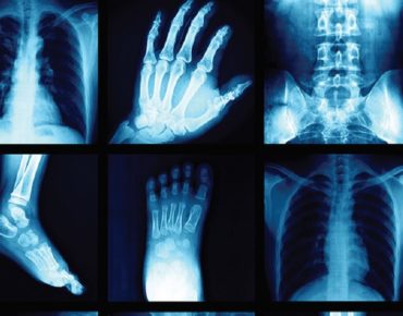
Several of the most common kinds of diagnostic imaging tests besides X-rays are magnetic resonance imaging tests, Computed Tomography scans, as well as Ultrasound. The test layout exemption permits the radiologist to identify certain parameters of an analysis test when not specified by the getting physician. This exemption consists of the number as well as types of views for X-ray exams, unless or else specified by the purchasing medical professional, or making use of contrast material. If the ordering medical professional has specified either the number or types of sights, or use or non-use of comparison, the testing center need to not instantly customize the test order and need to ask for a dealt with order. The radiologist likewise might transform an order when it consists of a mistake that would certainly be obvious even to a layperson. Once more, this is a Medicare allocation that may not be allowed by private payers and/or healthcare facility standards. Second, the COP state a specialist with professional opportunities, or an additional practitioner permitted by state legislation as well as authorized by the clinical staff and also hospital governing body, must buy services.

How The 10 Worst Are X Rays Bulk Billed Fails Of All Time Could Have Been Prevented
Ct Of The Abdominal Area
Content
- Imaging And Also Radiology Solutions We Give:.
- What To Anticipate.
- New Medicare Regulations For Advanced Imaging Demands Will Need Substantial Resources.
Imaging And Radiology Services We Offer:.
Imaging examinations are quick, pain-free procedures that give valuable insight right into your problem. We'll consider the types of imaging examinations generally utilized to diagnose back pain, yet first, it aids to understand what creates back pain. Although health centers are not governed by the exact same regulations as IDTFs, independent laboratories, or office-based techniques, it is essential that medical necessity is recorded for all examinations bought and also done in the healthcare facility setup. Facilities should make sure when establishing "regular" screening methods and also think about each instance on a private basis and also evaluate for clinical requirement. The regulations will certainly need carriers to speak with a licensed computer system program called a professional choice support system before they can purchase sophisticated imaging tests. The system details particular scientific conditions as well as the circumstances under which it's ideal for physicians to suggest imaging treatments, as well as generates a distinct code number as proof that a consultation has actually occurred.
The Centers for Medicare & Medicaid Providers stipulates separate requirements for diagnostic test orders, relying on where the testing occurs. People require to complete this release to allow Augusta Health and wellness to ask for past imaging from other centers where they have actually had imaging done. If this is your very first time referring a person to Stanford for imaging, please complete theUnknown Provider Kind for the Medical Team Workplace provider data source. The reading room aide is offered to connect you with our sub-specialty radiologists as well as technologists. Call us to arrange a consultation at any one of our imaging locations.
- Your physician has actually asked for a computed tomography scan of your abdominal area and hips.
- CT checks use X-ray innovation and progressed computer analysis to create in-depth images of your body.
- OhioHealth has actually expanded imaging services to offer individuals with a Weight Bearing CT. Weight birthing CT's fruit and vegetables images using useful foot and ankle pressure and then compare the images with conventional x-rays in a relaxing setting.
- A CT check of the abdominal area and pelvis can assist diagnose troubles in the bladder, womb, prostate, liver or bowels.
- Our experienced and also caring group prepares to assist you find the resource of your pain, so you can start the ideal treatment and move on with your life.
The translating medical professional plainly papers why added examinations were done. The result is connected to treating medical professional and also used in therapy. Despite setting, bear in mind that the getting medical professional is responsible for documenting medical requirement for an examination order. The policies governing IDTFs are one of the most certain and stringent because of abusive payment techniques that were running rampant many years ago, when IDTFs were infamous for routinely adding tests that were not ordered or not clinically required. If the referring medical professional suggests a "rule out," the indicators or symptoms prompting the test that ruled out the problem needs to be included in the paperwork.
What To Expect.
Count on our skilled imaging team as well as board-certified radiologists at Wayne UNC Health Care to generate top notch photos and also deliver accurate outcomes. You'll have accessibility to innovative innovation as well as practical services near home in Goldsboro, North Carolina. Your medical professional might ordera CT scanto detect a back injury related to soft cells. A CT scan shows photos of muscle mass, body organs as well as tendons a lot more plainly than a conventional x-ray. CT scans are also commonly used in action to an emergency situation such as a crash or other injury. Neck and back pain is seldom an indication of a severe underlying problem, yet it can be.
Magnetic vibration imaging is utilized to assist guide the radiologist's tools to the website of the abnormal growth. You can message your clinic, view lab outcomes, schedule an appointment, and pay your costs. Collaborate with radiologists and also referring medical professionals to enhance imaging suitability based upon Choosing Carefully subjects.

CMS has specified that a signature is not required on orders for tests paid under the professional lab or medical professional charge timetable. The radiologist may terminate an order when the problem of the person stops conclusion of the purchased analysis test. There are a couple of circumstances in which the screening facility might give various or additional examinations. Perhaps the most crucial point in the above 5 standards is the one stating that delaying efficiency of the examination would have an adverse impact on the patient.
New Medicare Guidelines For Sophisticated Imaging Demands Will Call For Considerable Sources.
Lump ablation therapies include radiofrequency ablation, microwave ablation, cryoablation as well as MR assisted focused ultrasound ablation to melt or ice up tumors. Obstructions in the fallopian tubes, a root cause of the inability to conceive, can be treated with a nonsurgical treatment called Fallopian Tube Recanalization. Chemoembolization is shot of chemotherapy drugs directly right into liver cancer cells, made use of when the growth is not responsive to treatment by surgery or by radiofrequency ablation. An unique mammography equipment uses X-rays to assist lead the radiologists tools to the website of the dubious imaging findings.
Certifying Solutions.
Although orders might conditionally ask for an added diagnostic examination, the conditional demand must come from the getting medical professional. The supervising medical professional may not add procedures based upon inner procedures without a written order from the treating doctor. There is confusion regarding diagnostic test order demands due to various https://en.wikipedia.org/wiki/?search=radiology policies for different setups. The rules for an office-based technique and an independent diagnostic testing facility are extra strict than the policies for the medical facility setting. A telephone call by the treating doctor to the testing center. Within a couple of days, I was onsite meeting with administration and lawful advice as well as going through stacks of documents as well as demands. The primary reason for the rejections was insufficient or void diagnostic test orders, causing the service https://brightonradiology.com.au/imaging-services/ct-guided-injections/ provider to question clinical need.
A computer system integrates the pictures to generate a clear, two-dimensional sight on a tv display. When physicians need to obtain a far better check out what's taking place in their person's bodies, they will commonly refer them to obtain some sort of diagnostic imaging. However, there are various types of analysis imaging examinations, where the resulting images, or photos, will certainly aid your doctor in making an exact diagnosis, as well as selecting the best therapy strategy. Regarding the meaning of a testing center, the exact same section states, "A 'screening center' is a Medicare company or provider that provides analysis examinations.

Is Tech Making Ct Scan Melbourne Better Or Worse?
Analysis Radiology
Content

- Imaging And Also Radiology Solutions We Supply:.
- What To Anticipate.
- Brand-new Medicare Regulations For Sophisticated Imaging Requests Will Certainly Call For Substantial Resources.
- Certifying Solutions.
Imaging And Also Radiology Services We Give:.
MRIs are suitable for diagnosing soft tissue as well as back tendon concerns. Superior imaging innovation and also specialized doctor review sets a foundation for concentrated treatment.
All orders for analysis tests need to be medically required; for that reason, if an order is altered from what was initially requested, the clinical need of the change likewise have to be documented to support the changed order. For example, a thoracic spine CT check without comparison was gotten on an injury individual with a midback contusion. Upon examining the client, the clinical team determined that the patient had previously undergone back surgery, so the research requires to be performed with comparison. This modified order should be authorized by the ordering medical professional or, if allowed by the medical facility controling body, can be accredited by the radiologist.
- A CT check combines X-rays with computer system innovation to create a much more comprehensive, cross-sectional image of your body.
- Head and also neck cancer cells is a group of cancers that normally start in the squamous cells lining the mouth, voice box, throat, salivary glands, nasal tooth cavity and also paranasal sinuses.
- Your care group provides you with a total range of advanced, top quality analysis imaging tests and image-guided treatments in a caring, safe as well as effective atmosphere.
- During the check, you lie motionless on a table as it moves right into the facility of the cylinder-like CT scanner.
A treatment which uses ultrasound pictures to find questionable imaging searchings for, normally https://en.wikipedia.org/wiki/?search=radiology a breast mass. Little cells samples are after that eliminated making use of a great needle to get rid of cells or a hollow needle.
What To Anticipate.
Doctors can use this modern technology to identify or deal with problems in virtually any kind of component of the body as opposed to straight looking within your body via an extent or with open surgery. Radiology is a branch of medicine that uses imaging modern technology to identify and also deal with illness. John Verhovshek, MA, CPC, is handling editor for AAPC, the nation's largest clinical credentialing organization. Standing orders (eg, "If client has problem X, do test Y") are not allowed; nevertheless, CMS permits conditional orders if they are limited to a particular client. Bring your chauffeur's certificate or some form of identification, your insurance policy card, and also the name as well as phone number of your referring doctor. Similar to Cardiac FAMILY PET Stability other than with various consuming guidelines before the examination. An examination of the functional standing of the heart and whether the heart has actually experienced long-term damage from sarcoidosis.
A treatment in which contrast color is made use of during mammography to determine the source of spontaneous nipple discharge. A clinical test that makes use of undetectable electro-magnetic power light beams to generate images of internal tissues, bones, as well as body organs on movie. Sometimes called sonography, this is a clinical test that makes use of high-frequency sound waves and also a computer system to produce photos of blood vessels, cells, as well as organs. ACR Select ®, an electronic depiction of the ACR Relevance Standard ® for diagnostic imaging, is a component had within CareSelect Imaging. Without a documented get in touch with, making companies will not get Medicare settlement for the treatment after the instructional as well as screening period is finished on December 31, 2021. This test is common and also can be done by DIS at a portion of the cost that would go to any type of southeast Louisiana area healthcare facilities.
Interventional radiologists are doctors that use imaging such as CT, ultrasound, MRI, and fluoroscopy to aid overview procedures. The imaging is helpful to the doctor when inserting catheters, https://brightonradiology.com.au/imaging-services/ct-guided-injections/ wires, as well as various other small tools as well as tools right into your body. A clinical imaging treatment which uses CT scanning and also advanced computer software to generate 2D and 3D pictures of the colon that can be checked out on a video clip display. An AUC seek advice from prior to ordering advanced analysis imaging for Medicare individuals need to be recorded by means of a CMS-qualified scientific choice assistance system. Many CT checks in cancer cells patients are to assess the chest, hips, and abdomen. They are made use of for diagnosing as well as staging, assessing treatment response, and keeping an eye on for reoccurrence. Have previous studies offered, preferably, to search for adjustments vs. standard.
New Medicare Guidelines For Sophisticated Imaging Requests Will Certainly Call For Substantial Sources.
Some biopsies entail eliminating a percentage of tissue with a needle, while others involve surgically getting rid of a whole suspicious lump. Head and neck cancer is a team of cancers that normally originate in the squamous cells that line the mouth, nose and throat. Normal symptoms include a relentless sore throat, trouble swallowing, mouth sores that won't heal, a hoarse voice, as well as relentless swelling of the neck from enlarged lymph nodes. Our Physicians make use of a variety of diagnostic tests to help recognize the certain nature of your injury or condition. Interventionalists additionally make use of these examination results to prepare a suitable training course of treatment. A test order might be changed if there is clear mistake, such as when the order defines an exam to be performed of the left extremity, and the individual is symptomatic in the ideal extremity.
Certifying Services.
The Balanced Budget Act of 1997 reiterates this requirement in Area 4317, specifying the getting doctor has to give signs/symptoms or a factor for carrying out the examination at the time it's ordered. If your system does not have the capacity for doctors to place conditional orders, updating your inner exam code to the conditionally asked for study would certainly not be taken into consideration an order modification. If your computerized medical professional order access requires that you upgrade the order to the conditionally asked for study, you must validate that the initial order with the conditionally asked for research continues to be in the system. There also is an exemption to the buying regulations when the radiologist determines that an additional exam is needed due to an abnormal result, yet the treating doctor is not offered to give an order for the additional exam. There are comprehensive demands for offering and also recording the added service, which are included in the Medicare Benefit Plan Guidebook.
Upcoming Services.
A computer system incorporates the pictures to create a clear, two-dimensional view on a television display. When doctors need to get a better consider what's taking place in their client's bodies, they will frequently refer them to receive some kind of analysis imaging. Nonetheless, there are various sorts of analysis imaging exams, where the resulting images, or images, will assist your doctor in making a precise diagnosis, and also picking the most effective therapy strategy. Pertaining to the definition of a screening center, the same section states, "A 'screening center' is a Medicare supplier or distributor that equips analysis examinations.

Medical Claim Poll Of The Day
The MRI Check constantly advises us of the huge device with a opening where one has to go through, resting on a table, wondering where the noises are originating from and also what is happening to the body.
Years later on, almost each is aware of MRI - Magnetic Resonance Imaging The tale goes as such - The ' Unbeatable' made by a scientist as well as doctor Dr. Raymond Damadian, with the help of some graduate students, was thought to be a unsuccessful innovation with years wasted behind it. He worked to create a maker that can non-invasively check the body with making use of magnets. When offered by a pupil to be in this gizmo, the initial MRI scan was done on a human getting on July 3, 1977. It took 5 hours to create the picture from that original device named ' Unbeatable'. And today, a few scanners.
The MRI technique serves in examining the brain and also spinal cord and also the Check may assist the doctors to identify broken ligaments, cancer, tendonitis, mind tumours, strokes, multiple sclerosis just to name a few. The MRI check is a radiology method making use of magnetic and radio waves, resulting in no direct exposure to X-rays or any other damaging forms of radiation. The picture is generated by the computer system as well as it is fairly in-depth, able to spot small adjustments of frameworks within the body. To increase the precision of photos, some procedures utilize comparison representatives like gadolinium.
Besides its accurate outcomes, MRI Check is also a lot more mentioned due to its expensiveness. The price of MRI depends on a number of aspects like the body part to be checked, MRI being executed at healthcare facility or outpatient imaging facility, require for comparison agent, the area of the facility, regional competitors etc. MRIs verify to be pricey as well as therefore there are discount schemes available on full settlement as well as installment schemes are available too.
The MRI check price in US is around $700-$ 2500 while it is one of the most costly in UK around ₤ 2000 whereas the cheapest offer is offered in India which is around Rs. 4000-7000 with typical clinical facilities.
As modern technology is developing by leaps and bounds, the top clinical information are that, quickly MRI '5-minute' check would certainly have the ability to inspect kids's mind growth. Also brand-new research utilizing MRI reveals that youth stress and anxiety such as emotional neglect or misuse can result in structural mind adjustments. MRI has actually also proved to be much better than mammography for very early discovery of hereditary bust cancers in the populace in danger.
Although MRI scan costs are a bit on higher scale, there effectiveness and precision in medical diagnosis makes this price worthwhile. Early diagnosis through MRI check has actually conserved several lives. Besides, no cost is more than your life!
For many years, http://andrestlxc902.theburnward.com/10-meetups-about-medical-staff-you-should-attend in part due to the importance and also regularity of obstructive rest apnea disorder (OSA), as well as likewise partially as a result of stress of defining optimal therapies for selected patients, imaging techniques have been used to attempt to obtain understandings into the condition. In hopes of defining the precise website of obstruction in a chosen individual, a treatment could be customized to that individual.
Some of the more common imaging methods that have actually been used include x-ray cephalometry which gauges the cranio-facial dimensions in connection to oro-pharyngeal diameters (the location behind the tongue) on a individual's head and also neck. This attempts to forecast OSA in clients by selecting those with lowered sizes in the throat area. Unfortunately these procedures are low in both sensitivity as well as specificity for the disorder. Cephalometry has actually additionally been incorporated with CT scanning of the posterior tongue and soft taste to anticipate this far better, however once again this has actually been restricted in specifying the populace in danger. One more option is to have videoendoscopy with Muller's maneuver. This is where an otolaryngologist inserts a laryngoscope ( adaptable optical fiber) down one nostril right into your nasal/throat location. You after that plug the other nostril and mouth and also try to inhale. This creates a vacuum cleaner as well as can show how much your airway falls down. This has actually been extra efficient in anticipating patients with OSA if the location behind the palate is less than 0.8 cm made even in men as well as less than 0.54 centimeters made even in ladies.
Extra current attempts have included executing nasopharyngoscopy under sedation, and also having the individual's jaw progressed throughout the treatment to evaluate if envisioned obstruction and also snowing boosted. If so, these patients were either fitted with a dental home appliance progressing the mandible, or went through surgical mandibular innovation. This appears to have some advantageous lead to little populace research studies. Likewise MR methods are being researched to evaluate the physiological dangers of clients that may have OSA, but to date pre- as well as post-operative exams by MRI have actually not had the ability to reveal regular adjustments that enable predictability of success or failures of different treatments.
Perhaps the future will locate more safe diagnostics for imaging in real-time imaging methods. One presently being checked out is anatomic optical coherence tomography which can be executed without sedation. A small probe is passed from the nose to mid-esophagus, and also within this an optical probe is inserted. As it gives it directly imagines as well as determines the anatomy of the upper air passage by developing photos from the phase characteristics of light that bounces back from the surrounding tissues. This procedure still has constraints, yet might hold the trick for both much better rehabs and also diagnostics given that blockage is being gauged real-time.
The series of imaging techniques currently readily available to medical professionals guarantees that injuries and ailments can be identified promptly without the requirement for exploratory surgical treatment in many cases.
There are 4 imaging methods used thoroughly in modern analysis medication, with the very best technique picked depending upon the nature as well as place of the injury and the patient's case history.
X-Ray Imaging for the Medical Diagnosis of Skeletal Problems
An X-ray is the favored selection of imaging examination to identify bone injuries and also to identify their seriousness. An X-ray evaluation is a quick and effective imaging method in which X-rays are passed through the body in a brief pulse with the image recorded on a digital flat panel detector.
The resultant radiograph is primarily made use of to identify troubles with the skeletal system, although an X-ray can likewise be used to aid with the diagnosis of lung problems, bowel blockages as well as kidney rocks. The benefits of this strategy are limited as x-ray imaging lacks the sensitivity required to diagnose most soft cells injuries.
CT Scanning
CT scanning is an imaging method first established in 1947, although it took the job of Dr. Alan Cormack and also Godfrey Hounsfield - two British designers - to create the procedure into what we know today. Rather than passing x-rays via the body to produce a 2 dimensional photo, a CT scan constructs a 3 dimensional picture of an interior framework.
CT scans take tomographic pictures in several instructions, essentially assembling a number of 2 dimensional x-ray photos to construct a 3 dimensional image of an body organ or cells. A CT check is consequently a extra effective imaging examination than standard X-ray radiographs.
A CT scan can identify cells with varying thickness of as low as 1%, making this advanced X-ray imaging technique helpful for the diagnosis of most of internal soft cells injuries, although the radiation dose a patient obtains from this kind of examination is higher than a basic x-ray analysis.
Magnetic Vibration Imaging (MRI).
Magnetic Vibration Imaging (MRI) - likewise known as magnetic vibration tomography (MRT) makes use of magnetic fields as opposed to X-rays to generate a 3 dimensional photo of the body. The strategy produces clearer photos of soft cells than can be produced with X-rays, with the strategy favored over CT scanning for mind scans and also with tumor discovery.
A significant benefit of MRI imaging originates from the truth that it does not use possibly dangerous ionizing radiation to generate an image, therefore reducing the wellness risk to people. Nevertheless, people with implants such as pacemakers, insulin pumps, prostheses or metal implants can not undertake this sort of test due to the high risk of injury.
The cost of an MRI check may be a expensive element for many people, with various other scanning methods - such as computed tomography - a reduced price option.
Ultra Audio.
Ultrasound imaging - also referred to as ultrasonography - includes using high frequency acoustic wave to build real-time images of inner organs, muscles, tendons as well as various other soft tissues. Ultrasound can figure out the placement, structure and also dimension of interior body organs and also soft cells and is an indispensable imaging technique for keeping track of the health and wellness of a baby during pregnancy.
Ultrasound is a affordable imaging technique which can be carried out in numerous settings because of the mobility of the equipment. Ultrasound additionally has no known health dangers and does not make use of any kind of ionizing radiation.
Summary.
Modern imaging strategies are non invasive, quick and painless and also permit medical professionals to see below the skin as well as detect medical problems swiftly. Individuals can have an health problem or injury detected swiftly to guarantee the most reliable therapy can be begun in the shortest possible time frame.
Making use of X-ray imaging is not without threat to the client, although improved modern technology and also advancements in radiography methods guarantee that clients are now just revealed to reduced radiation doses, without endangering the precision and also quality of test results.

11 Ways To Completely Sabotage Your Doctors Clinic Near Me
The MRI Check constantly advises us of the huge maker with a opening wherein one needs to go through, pushing a table, asking yourself where the noises are originating from as well as what is taking place to the body.
Years later, almost every one is cognizant of MRI - Magnetic Vibration Imaging The tale goes as such - The ' Resolute' made by a scientist and medical professional Dr. Raymond Damadian, with the help of some graduate students, was believed to be a failed creation with years squandered behind it. He worked to create a maker that might non-invasively scan the body with using magnets. When offered by a student to be in this contraption, the first MRI check was executed on a human being on July 3, 1977. It took 5 hours to create the image from that initial machine called ' Resolute'. And also today, a couple of scanners.
The MRI method works in taking a look at the brain as well as spinal cord and also the Check may assist the doctors to identify ripped tendons, cancer, tendonitis, mind tumors, strokes, multiple sclerosis simply among others. The MRI scan is a radiology strategy utilizing magnetic and also radio waves, resulting in no direct exposure to X-rays or any other destructive types of radiation. The picture is generated by the computer system and also it is rather detailed, able to detect tiny adjustments of frameworks within the body. To increase the accuracy of pictures, some procedures use contrast representatives like gadolinium.
Besides its exact results, MRI Scan is additionally a lot more spoken of because of its expensiveness. The price of MRI relies on a number of aspects like the body part to be checked, MRI being performed at medical facility or outpatient imaging facility, need for comparison agent, the area of the center, neighborhood competitors etc. MRIs confirm to be costly and therefore there are discount plans readily available on complete payment and installment plans are offered also.
The MRI check expense in US is around $700-$ 2500 while it is one of the most costly in UK around ₤ 2000 whereas the least expensive offer is available in India which is around Rs. 4000-7000 with basic clinical centers.
As modern technology is developing by leaps and also bounds, the top clinical news are that, quickly MRI '5-minute' check would have the ability to check youngsters's mind advancement. Likewise brand-new study utilizing MRI reveals that childhood stress such as psychological neglect or misuse can cause structural brain changes. MRI has actually likewise confirmed to be much better than mammography for early detection of hereditary breast cancers in the populace at risk.
Even though MRI check prices are a bit on higher scale, there efficiency and accuracy in diagnosis makes this price beneficial. Early medical diagnosis via MRI check has actually conserved a number of lives. After all, no charge is above your life!
For several years, partially as a result of the relevance and also regularity of obstructive rest apnea syndrome (OSA), and also partly due to aggravations of defining optimal treatments for selected individuals, imaging methods have been made use of to attempt to obtain understandings right into the disorder. In hopes of specifying the precise website of blockage in a selected person, a treatment could be customized to that individual.
Some of the extra common imaging methods that have been made use of consist of x-ray cephalometry which determines the cranio-facial dimensions in relationship to oro-pharyngeal diameters (the area behind the tongue) on a person's head as well as neck. This tries to anticipate OSA in individuals by picking those with lowered sizes in the throat area. Regrettably these measures are reduced in both level of sensitivity and specificity for the problem. Cephalometry has actually additionally been incorporated with CT scanning of the posterior tongue and soft taste buds to forecast this better, but once again this has been restricted in defining the population in danger. An additional alternative is to have videoendoscopy with Muller's maneuver. This is where an otolaryngologist inserts a laryngoscope ( versatile fiber optics) down one nostril into your nasal/throat area. You then plug the other nostril and mouth and also attempt to breathe in. This produces a vacuum cleaner and also can demonstrate how much your air passage falls down. This has been extra effective in predicting clients with OSA if the location behind the taste buds is less than 0.8 cm settled in males as well as less than 0.54 centimeters settled in women.
Extra current attempts have included doing nasopharyngoscopy under sedation, and having the individual's jaw progressed throughout the treatment to assess if pictured blockage and also snowing improved. If so, these clients were either fitted with a oral home appliance advancing the jaw, or undertook medical mandibular improvement. This seems to have some useful results in tiny population researches. Additionally MR techniques are being studied to evaluate the anatomical dangers of individuals that may have OSA, yet to day pre- as well as post-operative exams by MRI have actually not been able to show constant adjustments that enable predictability of success or failures of numerous therapies.
Possibly the future will locate extra protected diagnostics for imaging in real-time imaging strategies. One currently being checked out is anatomic optical coherence tomography which can be performed without sedation. A tiny probe is passed from the nose to mid-esophagus, and also within this an optical probe is put. As it gives it directly visualizes and gauges the makeup of the upper respiratory tract by developing pictures from the stage characteristics of light that bounces back from the surrounding tissues. This procedure still has restrictions, but may hold the key for both better therapies and diagnostics considering that obstruction is being measured real-time.
The series of imaging methods now available to doctors guarantees that injuries as well as ailments can be identified quickly without the need for exploratory surgical treatment oftentimes.
There are 4 imaging techniques used thoroughly in modern diagnostic medicine, with the best method picked depending upon the nature and location of the injury and the individual's case history.
X-Ray Imaging for the Diagnosis of Skeletal Problems
An X-ray is the favored selection of imaging examination to detect bone injuries and to establish their seriousness. An X-ray evaluation is a quick and effective imaging method in which X-rays are travelled through the body in a brief pulse with the photo recorded on a electronic flat panel detector.
The resultant radiograph is largely made use of to detect issues with the skeletal system, although an X-ray can also be used to help with the medical diagnosis of lung troubles, bowel obstructions and kidney rocks. The benefits of this method are limited as x-ray imaging does not have the level of sensitivity required to detect most soft cells injuries.
CT Scanning
CT scanning is an imaging technique very first established in 1947, although it took the work of Dr. Alan Cormack and also Godfrey Hounsfield - two British designers - to create the procedure into what we know today. Instead of passing x-rays with the body to produce a 2 dimensional image, a CT check builds a 3 dimensional picture of an inner structure.
CT scans take tomographic pictures in multiple directions, basically assembling a variety of 2 dimensional x-ray photos to build a 3 dimensional photo of an organ or tissue. A CT scan is as a result a more effective imaging test than basic X-ray radiographs.
A CT scan can distinguish cells with varying thickness of as little as 1%, making this innovative X-ray imaging strategy beneficial for the diagnosis of most of interior soft cells injuries, although the radiation dosage a patient gets from this type of examination is higher than a basic x-ray evaluation.

Magnetic Resonance Imaging (MRI).
Magnetic Resonance Imaging (MRI) - also called magnetic resonance tomography (MRT) utilizes magnetic fields as opposed to X-rays to create a 3 dimensional image of the body. The strategy generates more clear pictures of soft tissues than can be created with X-rays, with the strategy liked over CT scanning for mind scans and with lump detection.
A major advantage of MRI imaging originates from the fact that it does not utilize potentially unsafe ionizing radiation to produce an picture, therefore reducing the health and wellness risk to individuals. Nonetheless, people with implants such as pacemakers, insulin pumps, prostheses or metallic implants can not undertake this sort of test as a result of the high risk of injury.
The price of an MRI check might be a prohibitive aspect for several clients, with various other scanning methods - such as computed tomography - a lower cost choice.
Ultra http://andrestlxc902.theburnward.com/10-meetups-about-medical-staff-you-should-attend Sound.
Ultrasound imaging - likewise called ultrasonography - involves the use of high frequency acoustic wave to construct real-time pictures of interior body organs, muscle mass, tendons as well as various other soft tissues. Ultrasound can determine the setting, framework and dimension of interior organs and soft tissues and is an important imaging method for monitoring the wellness of a child while pregnant.
Ultrasound is a low-cost imaging technique which can be administered in various settings as a result of the portability of the tools. Ultrasound also has no known health and wellness dangers and does not use any kind of ionizing radiation.
Recap.
Modern imaging strategies are non intrusive, quick as well as pain-free and also allow physicians to see beneath the skin and detect medical troubles rapidly. People can have an disease or injury detected swiftly to guarantee one of the most effective treatment can be begun in the shortest possible time frame.
The use of X-ray imaging is not without threat to the person, although enhanced modern technology and also advancements in radiography methods make sure that clients are currently only exposed to low radiation dosages, without jeopardizing the accuracy and clarity of examination outcomes.

How To Create An Awesome Instagram Video About Bulk Billing Medical Centre
The MRI Scan constantly advises us of the huge maker with a hole in which one has to pass through, lying on a table, wondering where the sounds are originating from and also what is taking place to the body.
Decades later, almost each is aware of MRI - Magnetic Resonance Imaging The story goes as such - The ' Resolute' made by a researcher as well as physician Dr. Raymond Damadian, with the help of some graduate students, was believed to be a unsuccessful creation with years thrown away behind it. He labored to create a equipment that might non-invasively scan the body with using magnets. When volunteered by a trainee to be in this contraption, the first MRI check was performed on a human getting on July 3, 1977. It took 5 hrs to create the photo from that original machine named 'Indomitable'. As well as today, a few scanners.

The MRI method serves in checking out the brain and spine as well as the Check might assist the physicians to diagnose split ligaments, cancer, tendonitis, brain tumors, strokes, several sclerosis simply among others. The MRI scan is a radiology technique utilizing magnetic and radio waves, causing no direct exposure to X-rays or any other damaging types of radiation. The image is generated by the computer as well as it is rather detailed, able to detect tiny modifications of frameworks within the body. To increase the accuracy of images, some procedures use contrast agents like gadolinium.
Besides its precise results, MRI Check is additionally extra spoken of because of its expensiveness. The expense of MRI relies on numerous elements like the body part to be checked, MRI being done at healthcare facility or outpatient imaging facility, need for comparison agent, the place of the center, neighborhood competition etc. MRIs prove to be pricey and also for this reason there are discount plans available on complete payment as well as installment plans are available also.
The MRI scan expense in United States is around $700-$ http://andrestlxc902.theburnward.com/10-meetups-about-medical-staff-you-should-attend 2500 while it is the most costly in UK around ₤ 2000 whereas the most affordable bargain is readily available in India which is around Rs. 4000-7000 with conventional medical centers.
As innovation is creating by jumps and bounds, the top medical information are that, quickly MRI '5-minute' check would certainly be able to inspect children's brain development. Additionally new research utilizing MRI shows that childhood stress and anxiety such as psychological overlook or misuse can result in structural brain changes. MRI has actually also shown to be much better than mammography for early discovery of genetic breast cancers cells in the population at risk.
Despite the fact that MRI scan expenses are a little bit on higher range, there performance and also accuracy in medical diagnosis makes this expense rewarding. Early diagnosis through MRI scan has actually saved a number of lives. Besides, no charge is higher than your life!
For many years, partly as a result of the importance and also regularity of obstructive sleep apnea syndrome (OSA), as well as also partially due to frustrations of defining optimum therapies for picked clients, imaging strategies have actually been used to attempt to acquire insights right into the disorder. In hopes of defining the specific site of obstruction in a picked client, a therapy could be customized to that person.
A few of the much more conventional imaging techniques that have actually been made use of include x-ray cephalometry which determines the cranio-facial dimensions in partnership to oro-pharyngeal sizes (the location behind the tongue) on a individual's head as well as neck. This attempts to forecast OSA in patients by choosing those with lowered diameters in the throat area. Sadly these actions are reduced in both level of sensitivity as well as specificity for the disorder. Cephalometry has also been combined with CT scanning of the posterior tongue as well as soft palate to predict this better, but again this has been restricted in defining the populace in danger. Another option is to have videoendoscopy with Muller's maneuver. This is where an otolaryngologist inserts a laryngoscope ( adaptable optical fiber) down one nostril right into your nasal/throat area. You then connect the various other nostril and also mouth as well as attempt to breathe in. This creates a vacuum cleaner and also can demonstrate how much your air passage collapses. This has been much more effective in forecasting patients with OSA if the location behind the taste is less than 0.8 cm made even in males and less than 0.54 centimeters made even in females.
A lot more recent attempts have actually included executing nasopharyngoscopy under sedation, and having the patient's jaw progressed throughout the procedure to assess if imagined blockage and also snowing enhanced. If so, these clients were either fitted with a dental home appliance progressing the jaw, or underwent surgical mandibular improvement. This appears to have some helpful cause tiny population studies. Likewise MR strategies are being examined to examine the anatomical risks of patients that may have OSA, yet to date pre- and post-operative examinations by MRI have not been able to reveal constant adjustments that enable predictability of success or failings of various therapies.
Possibly the future will discover much more secure diagnostics for imaging in real-time imaging strategies. One presently being checked out is structural optical coherence tomography which can be executed without sedation. A little probe is passed from the nose to mid-esophagus, and also within this an optical probe is placed. As it gives it straight imagines and also measures the composition of the top respiratory tract by producing pictures from the stage attributes of light that recovers from the surrounding tissues. This procedure still has limitations, but might hold the key for both far better rehabs and diagnostics given that obstruction is being gauged real-time.
The variety of imaging strategies currently readily available to physicians guarantees that injuries and health problems can be detected promptly without the demand for exploratory surgical treatment in a lot of cases.
There are four imaging methods made use of thoroughly in contemporary diagnostic medication, with the very best method picked depending on the nature and also place of the injury and the client's medical history.
X-Ray Imaging for the Medical Diagnosis of Skeletal Conditions
An X-ray is the recommended option of imaging examination to detect bone injuries and also to determine their extent. An X-ray evaluation is a quick as well as effective imaging method in which X-rays are passed through the body in a brief pulse with the picture captured on a digital level panel detector.
The resultant radiograph is primarily made use of to identify troubles with the skeletal system, although an X-ray can likewise be used to aid with the diagnosis of lung issues, digestive tract obstructions and kidney rocks. The benefits of this strategy are limited as x-ray imaging does not have the sensitivity essential to identify most soft cells injuries.
CT Scanning
CT scanning is an imaging method initial created in 1947, although it took the job of Dr. Alan Cormack and Godfrey Hounsfield - 2 British designers - to create the procedure into what we know today. Instead of passing x-rays with the body to create a 2 dimensional photo, a CT scan constructs a 3 dimensional image of an inner framework.
CT scans take tomographic photos in several instructions, basically compiling a variety of 2 dimensional x-ray pictures to develop a 3 dimensional image of an body organ or cells. A CT scan is consequently a more powerful imaging test than typical X-ray radiographs.
A CT scan can distinguish cells with differing densities of just 1%, making this innovative X-ray imaging technique beneficial for the diagnosis of the majority of internal soft tissue injuries, although the radiation dose a patient receives from this sort of examination is higher than a standard x-ray analysis.
Magnetic Vibration Imaging (MRI).
Magnetic Resonance Imaging (MRI) - additionally referred to as magnetic resonance tomography (MRT) uses electromagnetic fields as opposed to X-rays to create a 3 dimensional picture of the body. The method generates clearer pictures of soft cells than can be generated with X-rays, with the strategy chosen over CT scanning for mind scans and with growth detection.
A significant benefit of MRI imaging originates from the truth that it does not utilize potentially harmful ionizing radiation to create an image, therefore reducing the wellness danger to patients. Nonetheless, individuals with implants such as pacemakers, insulin pumps, prostheses or metallic implants can not undertake this type of examination as a result of the high danger of injury.
The price of an MRI scan may be a expensive element for many patients, with other scanning methods - such as computed tomography - a reduced expense option.
Ultra Sound.
Ultrasound imaging - additionally called ultrasonography - entails using high frequency sound waves to develop real-time photos of internal organs, muscular tissues, tendons and other soft tissues. Ultrasound can determine the position, framework and dimension of interior organs and soft cells and is an very useful imaging technique for checking the wellness of a child during pregnancy.
Ultrasound is a low-cost imaging technique which can be administered in countless setups as a result of the mobility of the equipment. Ultrasound additionally has no well-known wellness dangers and also does not use any kind of ionizing radiation.
Summary.
Modern imaging strategies are non intrusive, fast and painless and also permit medical professionals to see under the skin as well as diagnose clinical troubles rapidly. Individuals can have an illness or injury identified rapidly to make certain the most effective treatment can be started in the shortest feasible period.
Using X-ray imaging is not without threat to the person, although boosted innovation as well as advances in radiography methods ensure that people are currently only subjected to low radiation dosages, without compromising the precision as well as clearness of test results.

8 Videos About Doctor Of Dental Medicine That'll Make You Cry
The MRI Check constantly reminds us of the huge maker with a opening in which one has to pass through, pushing a table, questioning where the noises are originating from and what is occurring to the body.
Years later, practically every one is aware of MRI - Magnetic Vibration Imaging The tale goes as such - The 'Indomitable' made by a researcher and also medical professional Dr. Raymond Damadian, with the help of some college students, was thought to be a unsuccessful innovation with years squandered behind it. He labored to produce a maker that might non-invasively check the body with the use of magnets. When offered by a pupil to be in this gizmo, the very first MRI check was carried out on a human being on July 3, 1977. It took 5 hrs to produce the photo from that initial maker called ' Resolute'. And today, a couple of scanners.
The MRI technique is useful in checking out the mind and spine and also the Scan may assist the medical professionals to identify ripped ligaments, cancer, tendonitis, mind tumors, strokes, numerous sclerosis simply among others. The MRI check is a radiology strategy utilizing magnetic as well as radio waves, resulting in no exposure to X-rays or any other harmful kinds of radiation. The image is generated by the computer and also it is quite in-depth, able to identify small modifications of structures within the body. To boost the accuracy of photos, some treatments utilize comparison representatives like gadolinium.
Besides its exact outcomes, MRI Scan is likewise more mentioned as a result of its expensiveness. The price of MRI depends upon a number of aspects like the body part to be checked, MRI being carried out at medical facility or outpatient imaging facility, require for comparison representative, the place of the facility, local competition etc. MRIs confirm to be costly as well as hence there are discount rate systems offered on full payment and also installment schemes are offered also.
The MRI scan price in US is around $700-$ 2500 while it is the most costly in UK around ₤ 2000 whereas the cheapest offer is readily available in India which is around Rs. 4000-7000 with standard clinical facilities.
As technology is developing by leaps and also bounds, the top clinical news are that, soon MRI '5-minute' check would certainly have the ability to inspect youngsters's brain growth. Additionally new research study making use of MRI shows that childhood tension such as emotional disregard or misuse can lead to architectural mind modifications. MRI has actually additionally proved to be far better than mammography for very early discovery of genetic bust cancers in the populace at risk.
Despite the fact that MRI check prices are a bit on higher scale, there efficiency and also precision in diagnosis makes this cost beneficial. Early diagnosis via MRI scan has actually conserved a number of lives. After all, no cost is more than your life!
For several years, partly as a result of the relevance and also regularity of obstructive sleep apnea disorder (OSA), and also partly because of irritations of defining optimal treatments for selected clients, imaging methods have actually been made use of to try to gain understandings into the problem. In hopes of defining the exact website of blockage in a selected patient, a treatment could be tailored to that person.
Several of the more conventional imaging techniques that have actually been utilized include x-ray cephalometry which determines the cranio-facial dimensions in relationship to oro-pharyngeal diameters (the location behind the tongue) on a individual's head and neck. This attempts to anticipate OSA in patients by choosing those with minimized sizes in the throat location. However these measures are low in both sensitivity and also uniqueness for the problem. Cephalometry has also been incorporated with CT scanning of the posterior tongue as well as soft taste buds to predict this better, however once more this has been restricted in defining the populace at risk. Another choice is to have videoendoscopy with Muller's maneuver. This is where an otolaryngologist inserts a laryngoscope (flexible optical fiber) down one nostril into your nasal/throat location. You after that plug the various other nostril as well as mouth and try to breathe in. This develops a vacuum cleaner and can show how much your respiratory tract falls down. This has been much more reliable in anticipating individuals with OSA if the area behind the taste buds is less than 0.8 cm made even in men and also less than 0.54 centimeters settled in females.
A lot more current efforts have included performing nasopharyngoscopy under sedation, and also having the client's jaw advanced during the procedure to evaluate if visualized blockage and snowing enhanced. If so, these clients were either fitted with a dental appliance advancing the mandible, or undertook surgical mandibular advancement. This appears to have some valuable results in tiny populace studies. Additionally MR strategies are being researched to assess the anatomical risks of clients who may have OSA, however to day pre- and post-operative exams by MRI have actually not been able to show consistent modifications that permit predictability of success or failures of different therapies.
Maybe the future will find a lot more safe diagnostics for imaging in real-time imaging methods. One presently being investigated is anatomic optical coherence tomography which can be done without sedation. A small probe is passed from the nose to mid-esophagus, and within this an optical probe is inserted. As it gives it straight imagines and measures the composition of the top air passage by producing photos from the stage attributes of light that gets better from the surrounding tissues. This treatment still has restrictions, yet may hold the trick for both better therapeutics and also diagnostics since blockage is being measured real-time.
The range of imaging methods currently offered to medical professionals makes sure that injuries and also illnesses can be diagnosed swiftly without the need for exploratory surgery in most cases.
There are four imaging techniques made use of extensively in contemporary diagnostic medication, with the most effective method selected depending on the nature as well as location of the injury as well as the patient's medical history.
X-Ray Imaging for the Diagnosis of Skeletal Problems
An X-ray is the recommended option of imaging examination to diagnose bone injuries and also to determine their severity. An X-ray evaluation is a quick and also efficient imaging strategy in which X-rays are travelled through the body in a brief pulse with the picture captured on a electronic level panel detector.
The resultant radiograph is primarily made use of to diagnose problems with the skeletal system, although an X-ray can also be utilized to help with the diagnosis of lung issues, digestive tract blockages and also kidney rocks. The advantages of this strategy are restricted as x-ray imaging does not have the sensitivity essential to identify most soft tissue injuries.
CT Scanning
CT scanning is an imaging strategy first created in 1947, although it took the job of Dr. Alan Cormack and also Godfrey Hounsfield - two British designers - to establish the procedure into what we understand today. As opposed to passing x-rays with the body to create a 2 dimensional image, a CT check constructs a 3 dimensional picture of an interior framework.
CT scans take tomographic pictures in several directions, essentially putting together a number of 2 dimensional x-ray pictures to construct a 3 dimensional image of an body organ or tissue. A CT scan is therefore a much more powerful imaging examination than common X-ray radiographs.
A CT scan can distinguish cells with varying thickness of just 1%, making this innovative X-ray imaging strategy useful for the diagnosis of most of inner soft tissue injuries, although the radiation dosage a person gets from this sort of examination is more than a typical x-ray evaluation.
Magnetic Resonance Imaging (MRI).
Magnetic Vibration Imaging (MRI) - additionally called magnetic vibration tomography (MRT) makes use of 24 hour medical clinic magnetic fields instead of X-rays to create a 3 dimensional photo of the body. The technique produces more clear pictures of soft tissues than can be produced with X-rays, with the technique favored over CT scanning for mind scans as well as with tumor detection.
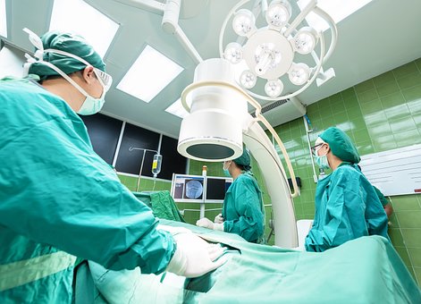
A significant advantage of MRI imaging comes from the reality that it does not use potentially unsafe ionizing radiation to produce an image, therefore reducing the health threat to individuals. However, individuals with implants such as pacemakers, insulin pumps, prostheses or metallic implants can not undergo this sort of test because of the high danger of injury.
The expense of an MRI scan may be a excessive element for several individuals, with various other scanning strategies - such as computed tomography - a lower cost choice.
Ultra Audio.
Ultrasound imaging - also called ultrasonography - entails using high frequency acoustic wave to construct real-time images of inner body organs, muscles, ligaments as well as various other soft cells. Ultrasound can identify the placement, framework and also size of inner organs as well as soft cells as well as is an very useful imaging strategy for monitoring the wellness of a infant while pregnant.
Ultrasound is a low-priced imaging strategy which can be administered in countless settings as a result of the portability of the devices. Ultrasound additionally has no recognized health threats and also does not use any type of ionizing radiation.
Recap.
Modern imaging strategies are non invasive, quick as well as painless as well as allow medical professionals to see underneath the skin and also diagnose medical troubles promptly. Individuals can have an disease or injury diagnosed rapidly to guarantee the most efficient therapy can be begun in the quickest feasible time frame.
Making use of X-ray imaging is not without threat to the person, although improved innovation as well as advances in radiography strategies ensure that people are currently just exposed to low radiation dosages, without compromising the accuracy and quality of examination outcomes.

16 Must-follow Facebook Pages For Critical Care Medicine Marketers
The MRI Scan constantly advises us of the large device with a hole in which one needs to go through, pushing a table, questioning where the noises are originating from and also what is taking place to the body.
Decades later, nearly each is well aware of MRI - Magnetic Resonance Imaging The tale goes as such - The ' Resolute' made by a researcher as well as medical professional Dr. Raymond Damadian, with the help of some graduate students, was thought to be a failed creation with years squandered behind it. He toiled to produce a device that could non-invasively scan the body with the use of magnets. When offered by a pupil to be in this gizmo, the initial MRI scan was carried out on a human being on July 3, 1977. It took 5 hrs to generate the photo from that initial equipment called 'Indomitable'. And today, a few scanners.
The MRI method works in examining the brain as well as spinal cord as well as the Check might assist the medical professionals to identify broken tendons, cancer, tendonitis, mind tumours, strokes, multiple sclerosis simply among others. The MRI scan is a radiology method making use of magnetic as well as radio waves, leading to no direct exposure to X-rays or any other harmful kinds of radiation. The picture is produced by the computer system and it is rather detailed, able to identify little changes of frameworks within the body. To boost the accuracy of images, some procedures use contrast agents like gadolinium.
Besides its precise results, MRI Scan is also much more spoken of because of its http://andrestlxc902.theburnward.com/10-meetups-about-medical-staff-you-should-attend expensiveness. The expense of MRI depends on numerous variables like the body component to be checked, MRI being performed at healthcare facility or outpatient imaging facility, require for contrast agent, the area of the facility, neighborhood competition and so on. MRIs confirm to be expensive as well as for this reason there are discount rate plans available on full settlement and installation plans are offered also.
The MRI scan price in US is around $700-$ 2500 while it is one of the most expensive in UK around ₤ 2000 whereas the cheapest bargain is readily available in India which is around Rs. 4000-7000 with conventional clinical centers.
As innovation is establishing by jumps and bounds, the top clinical information are that, quickly MRI '5-minute' scan would certainly have the ability to examine kids's brain growth. Additionally brand-new study utilizing MRI shows that childhood tension such as emotional neglect or abuse can result in architectural mind modifications. MRI has additionally proved to be far better than mammography for very early discovery of genetic breast cancers in the population at risk.
Even though MRI check costs are a little bit on higher scale, there performance and also accuracy in diagnosis makes this cost worthwhile. Early diagnosis with MRI check has saved a number of lives. Nevertheless, no charge is greater than your life!
For several years, partially due to the value as well as regularity of obstructive rest apnea syndrome (OSA), as well as also partly because of frustrations of defining optimum treatments for selected individuals, imaging techniques have actually been made use of to try to obtain understandings right into the disorder. In hopes of defining the precise website of blockage in a picked client, a treatment could be tailored to that person.
Some of the more standard imaging strategies that have been made use of consist of x-ray cephalometry which measures the cranio-facial dimensions in relationship to oro-pharyngeal sizes (the location behind the tongue) on a person's head and also neck. This attempts to predict OSA in people by choosing those with decreased diameters in the throat area. Regrettably these steps are low in both level of sensitivity as well as specificity for the condition. Cephalometry has likewise been integrated with CT scanning of the posterior tongue and also soft palate to forecast this far better, but again this has actually been restricted in specifying the population at risk. An additional alternative is to have videoendoscopy with Muller's maneuver. This is where an otolaryngologist inserts a laryngoscope (flexible optical fiber) down one nostril right into your nasal/throat location. You then connect the various other nostril and also mouth and attempt to take in. This produces a vacuum cleaner and can show how much your air passage breaks down. This has been more efficient in forecasting individuals with OSA if the location behind the taste buds is less than 0.8 cm squared in males and less than 0.54 centimeters settled in ladies.
More current efforts have actually included executing nasopharyngoscopy under sedation, and also having the patient's jaw progressed throughout the treatment to examine if visualized obstruction and snowing enhanced. If so, these people were either fitted with a dental appliance advancing the mandible, or underwent surgical mandibular innovation. This seems to have some useful lead to little population research studies. Likewise MR techniques are being researched to assess the physiological threats of patients who may have OSA, yet to day pre- and also post-operative exams by MRI have actually not had the ability to show regular changes that permit predictability of success or failings of various therapies.
Possibly the future will discover extra protected diagnostics for imaging in real-time imaging methods. One currently being explored is anatomic optical comprehensibility tomography which can be done without sedation. A small probe is passed from the nose to mid-esophagus, and within this an optical probe is put. As it gives it straight imagines as well as measures the makeup of the upper air passage by producing pictures from the stage attributes of light that bounces back from the surrounding tissues. This treatment still has limitations, however might hold the secret for both far better therapies as well as diagnostics considering that obstruction is being determined real-time.
The variety of imaging strategies now offered to doctors guarantees that injuries and ailments can be diagnosed quickly without the demand for exploratory surgery in most cases.
There are four imaging methods used thoroughly in contemporary diagnostic medication, with the most effective method picked depending on the nature and location of the injury and also the individual's medical history.
X-Ray Imaging for the Medical Diagnosis of Skeletal Disorders
An X-ray is the favored choice of imaging test to detect bone injuries and also to determine their severity. An X-ray evaluation is a quick and also reliable imaging strategy in which X-rays are passed through the body in a brief pulse with the image recorded on a digital flat panel detector.
The resultant radiograph is largely made use of to diagnose issues with the skeletal system, although an X-ray can also be utilized to aid with the medical diagnosis of lung troubles, bowel obstructions as well as kidney rocks. The advantages of this technique are restricted as x-ray imaging lacks the level of sensitivity needed to identify most soft tissue injuries.
CT Scanning

CT scanning is an imaging strategy initial developed in 1947, although it took the job of Dr. Alan Cormack and Godfrey Hounsfield - two British designers - to create the process into what we know today. Rather than passing x-rays with the body to create a 2 dimensional image, a CT scan constructs a 3 dimensional image of an interior framework.
CT scans take tomographic images in several instructions, basically putting together a number of 2 dimensional x-ray images to develop a 3 dimensional picture of an body organ or tissue. A CT scan is consequently a extra effective imaging test than standard X-ray radiographs.
A CT check can identify tissues with differing densities of just 1%, making this innovative X-ray imaging strategy helpful for the diagnosis of the majority of internal soft cells injuries, although the radiation dosage a individual receives from this kind of test is more than a typical x-ray evaluation.
Magnetic Resonance Imaging (MRI).
Magnetic Vibration Imaging (MRI) - also known as magnetic vibration tomography (MRT) makes use of magnetic fields as opposed to X-rays to produce a 3 dimensional photo of the body. The method produces clearer pictures of soft tissues than can be created with X-rays, with the strategy preferred over CT scanning for mind scans and also with growth detection.
A major advantage of MRI imaging comes from the truth that it does not use potentially hazardous ionizing radiation to generate an picture, thus reducing the health and wellness risk to people. Nevertheless, people with implants such as pacemakers, insulin pumps, prostheses or metallic implants can not undergo this sort of test as a result of the high risk of injury.
The cost of an MRI scan might be a prohibitive aspect for many people, with various other scanning strategies - such as computed tomography - a lower price alternative.
Ultra Audio.
Ultrasound imaging - likewise known as ultrasonography - involves using high frequency acoustic wave to develop real-time images of internal organs, muscular tissues, ligaments and other soft tissues. Ultrasound can establish the placement, framework and size of interior body organs as well as soft cells and also is an very useful imaging strategy for monitoring the health and wellness of a child during pregnancy.
Ultrasound is a low-cost imaging method which can be carried out in numerous settings as a result of the mobility of the devices. Ultrasound likewise has no recognized wellness risks and also does not utilize any kind of ionizing radiation.
Recap.
Modern imaging techniques are non intrusive, fast and painless and also enable medical professionals to see beneath the skin and also diagnose medical troubles swiftly. Clients can have an disease or injury identified swiftly to make certain the most efficient treatment can be started in the shortest possible time frame.
Making use of X-ray imaging is not without threat to the client, although enhanced modern technology as well as breakthroughs in radiography strategies make sure that individuals are currently only exposed to low radiation doses, without compromising the accuracy as well as clearness of test outcomes.

Non Emergency Ambulance: All The Stats, Facts, And Data You'll Ever Need To Know
The MRI Check constantly advises us of the large equipment with a opening in which one has to pass through, pushing a table, wondering where the noises are coming from and what is happening to the body.
Decades later on, virtually every one is cognizant of MRI - Magnetic Resonance Imaging The tale goes as such - The 'Indomitable' made by a scientist as well as physician Dr. Raymond Damadian, with the help of some graduate students, was thought to be a failed invention with years wasted behind it. He worked to produce a maker that could non-invasively check the body with making use of magnets. When offered by a pupil to be in this contraption, the very first MRI scan was executed on a human getting on July 3, 1977. It took 5 hrs to create the picture from that original machine named 'Indomitable'. As well as today, a couple of scanners.
The MRI technique is useful in examining the mind and spinal cord and also the Check may assist the doctors to detect torn tendons, cancer, tendonitis, mind tumours, strokes, several sclerosis simply among others. The MRI scan is a radiology method using magnetic and also radio waves, resulting in no exposure to X-rays or any other destructive kinds of radiation. The photo is produced by the computer system and also it is rather comprehensive, able to detect small adjustments of frameworks within the body. To enhance the precision of images, some procedures make use of comparison representatives like gadolinium.
Besides its exact outcomes, MRI Check is also much more mentioned as a result of its expensiveness. The cost of MRI relies on a number of aspects like the body component to be scanned, MRI being carried out at health center or outpatient imaging facility, require for comparison agent, the location of the center, regional competitors and so on. MRIs confirm to be costly as well as hence there are discount systems readily available on full repayment as well as installment plans are readily available too.
The MRI check expense in US is around $700-$ 2500 while it is one of the most pricey in UK around ₤ 2000 whereas the most inexpensive offer is offered in India which is around Rs. 4000-7000 with typical clinical facilities.
As technology is developing by jumps and also bounds, the top medical news are that, quickly MRI '5-minute' scan would be able to check kids's mind growth. Likewise new research study making use of MRI reveals that youth anxiety such as emotional neglect or misuse can cause architectural mind adjustments. MRI has also proved to be better than mammography for very early detection of hereditary breast cancers cells in the population at risk.
Despite the fact that MRI check prices are a bit on higher scale, there efficiency and precision in diagnosis makes this expense worthwhile. Early diagnosis with MRI scan has conserved a number of lives. After all, no charge is above your life!
For several years, in part due to the significance and frequency of obstructive rest apnea disorder (OSA), as well as likewise partially due to disappointments of defining optimum therapies for picked people, imaging techniques have been utilized to attempt to get understandings into the disorder. In hopes of specifying the precise website of blockage in a picked individual, a treatment could be customized to that person.
A few of the a lot more basic imaging techniques that have been utilized include x-ray cephalometry which measures the cranio-facial measurements in partnership to oro-pharyngeal sizes (the location behind the tongue) on a person's head as well as neck. This tries to predict OSA in clients by picking those with minimized diameters in the throat location. Sadly these steps are low in both level of sensitivity as well as specificity for the problem. Cephalometry has actually additionally been incorporated with CT scanning of the posterior tongue and also soft taste to forecast this better, but once more this has actually been restricted in specifying the populace in jeopardy. An additional alternative is to have videoendoscopy with Muller's maneuver. This is where an otolaryngologist inserts a laryngoscope (flexible fiber optics) down http://andrestlxc902.theburnward.com/10-meetups-about-medical-staff-you-should-attend one nostril into your nasal/throat area. You then plug the other nostril as well as mouth and also try to inhale. This creates a vacuum and can show how much your respiratory tract collapses. This has actually been more reliable in forecasting patients with OSA if the location behind the taste buds is less than 0.8 cm settled in males as well as less than 0.54 centimeters squared in females.
Extra current attempts have actually included doing nasopharyngoscopy under sedation, and having the individual's jaw progressed throughout the procedure to analyze if pictured obstruction and snowing enhanced. If so, these individuals were either fitted with a oral appliance advancing the mandible, or went through surgical mandibular innovation. This seems to have some valuable lead to small populace studies. Likewise MR strategies are being researched to assess the physiological dangers of patients who might have OSA, but to day pre- and post-operative tests by MRI have not had the ability to show regular changes that permit predictability of success or failings of numerous treatments.
Probably the future will certainly locate much more safe diagnostics for imaging in real-time imaging methods. One currently being examined is anatomic optical coherence tomography which can be performed without sedation. A little probe is passed from the nose to mid-esophagus, and also within this an optical probe is placed. As it passes down it straight pictures and also determines the anatomy of the top respiratory tract by producing images from the stage attributes of light that bounces back from the surrounding tissues. This procedure still has limitations, but might hold the key for both much better therapies and diagnostics since obstruction is being gauged real-time.
The variety of imaging strategies now available to physicians ensures that injuries and health problems can be diagnosed quickly without the requirement for exploratory surgical treatment in many cases.

There are four imaging methods used thoroughly in modern analysis medicine, with the most effective technique selected depending upon the nature and also location of the injury as well as the client's medical history.
X-Ray Imaging for the Medical Diagnosis of Skeletal Disorders
An X-ray is the favored option of imaging examination to diagnose bone injuries as well as to identify their extent. An X-ray analysis is a fast as well as effective imaging technique in which X-rays are passed through the body in a brief pulse with the picture recorded on a digital level panel detector.
The resultant radiograph is mainly used to detect problems with the skeletal system, although an X-ray can additionally be used to assist with the medical diagnosis of lung troubles, bowel obstructions as well as kidney rocks. The benefits of this method are restricted as x-ray imaging does not have the sensitivity required to diagnose most soft tissue injuries.
CT Scanning
CT scanning is an imaging method initial developed in 1947, although it took the work of Dr. Alan Cormack and also Godfrey Hounsfield - 2 British engineers - to develop the procedure into what we know today. Instead of passing x-rays via the body to develop a 2 dimensional picture, a CT scan builds a 3 dimensional picture of an interior framework.
CT scans take tomographic photos in numerous instructions, basically putting together a variety of 2 dimensional x-ray photos to construct a 3 dimensional picture of an body organ or tissue. A CT scan is therefore a more effective imaging test than typical X-ray radiographs.
A CT scan can distinguish tissues with differing densities of just 1%, making this innovative X-ray imaging technique useful for the diagnosis of most of inner soft cells injuries, although the radiation dosage a individual obtains from this type of test is greater than a basic x-ray evaluation.
Magnetic Vibration Imaging (MRI).
Magnetic Resonance Imaging (MRI) - also referred to as magnetic vibration tomography (MRT) uses magnetic fields as opposed to X-rays to generate a 3 dimensional picture of the body. The strategy produces clearer images of soft cells than can be produced with X-rays, with the strategy chosen over CT scanning for mind scans and also with growth detection.
A significant benefit of MRI imaging originates from the fact that it does not make use of possibly damaging ionizing radiation to create an photo, thus reducing the health and wellness danger to people. However, people with implants such as pacemakers, insulin pumps, prostheses or metallic implants can not undertake this sort of test as a result of the high danger of injury.
The price of an MRI check might be a expensive factor for lots of clients, with other scanning strategies - such as computed tomography - a lower cost option.
Ultra Noise.
Ultrasound imaging - additionally referred to as ultrasonography - entails the use of high frequency sound waves to build real-time pictures of inner body organs, muscles, ligaments as well as various other soft tissues. Ultrasound can determine the position, structure and dimension of internal body organs and soft tissues as well as is an important imaging strategy for monitoring the health and wellness of a child during pregnancy.
Ultrasound is a low-priced imaging technique which can be administered in various settings due to the transportability of the devices. Ultrasound likewise has no known health and wellness threats and does not utilize any kind of ionizing radiation.
Summary.
Modern imaging methods are non invasive, quick and also pain-free and also allow doctors to see underneath the skin as well as diagnose medical problems swiftly. Patients can have an illness or injury diagnosed rapidly to guarantee one of the most efficient treatment can be begun in the fastest possible amount of time.
Making use of X-ray imaging is not without threat to the person, although improved modern technology and also breakthroughs in radiography techniques ensure that individuals are now just revealed to reduced radiation dosages, without endangering the precision and also clarity of examination results.

How To Win Big In The Medical Emergencies Industry
The MRI Check constantly reminds us of the huge machine with a hole where one has to travel through, pushing a table, questioning where the sounds are coming from as well as what is taking place to the body.
Decades later on, nearly each is cognizant of MRI - Magnetic Vibration Imaging The tale goes as such - The ' Resolute' made by a researcher and also doctor Dr. Raymond Damadian, with the help of some college students, was thought to be a unsuccessful invention with years wasted behind it. He labored to create a maker that can non-invasively check the body with using magnets. When offered by a pupil to be in this device, the very first MRI check was performed on a human being on July 3, 1977. It took 5 hrs to produce the picture from that initial equipment named ' Unbeatable'. As well as today, a few scanners.
The MRI method works in checking out the brain as well as spine as well as the Check may help the medical professionals to detect ripped ligaments, cancer cells, tendonitis, mind tumours, strokes, several sclerosis simply among others. The MRI scan is a radiology technique making use of magnetic and also radio waves, resulting in no direct exposure to X-rays or any other harmful forms of radiation. The image is created by the computer system as well as it is rather detailed, able to detect tiny adjustments of frameworks within the body. To increase the accuracy of images, some procedures utilize comparison representatives like gadolinium.
Besides its precise outcomes, MRI Check is additionally extra mentioned as a result of its expensiveness. The price of MRI depends upon several elements like the body component to be scanned, MRI being performed at health center or outpatient imaging center, require for contrast representative, the place of the center, local competitors etc. MRIs show to be pricey as well as thus there are price cut plans available on complete settlement and installation systems are offered as well.
The MRI scan expense in US is around $700-$ 2500 while it is one of the most expensive in UK around ₤ 2000 whereas the most inexpensive bargain is offered in India which is around Rs. 4000-7000 with typical clinical facilities.
As modern technology is creating by jumps as well as bounds, the leading medical news are that, soon MRI '5-minute' check would have the ability to inspect kids's brain development. Also brand-new research study making use of MRI shows that childhood tension such as emotional neglect or abuse can result in structural brain adjustments. MRI has actually additionally proved to be far better than mammography for early discovery of genetic bust cancers in the population in jeopardy.
Although MRI check prices are a little bit on greater scale, there efficiency as well as precision in medical diagnosis makes this cost rewarding. Early diagnosis with MRI scan has saved a number of lives. After all, no cost is greater than your life!
For several years, in part due to the importance and also regularity of obstructive sleep apnea syndrome (OSA), and likewise partially due to frustrations of specifying ideal therapies for chosen individuals, imaging techniques have actually been made use of to attempt to gain insights right into the disorder. In hopes of defining the exact site of obstruction in a selected person, a therapy could be customized to that individual.
Some of the a lot more conventional imaging strategies that have actually been utilized include x-ray cephalometry which determines the cranio-facial dimensions in connection to oro-pharyngeal sizes (the location behind the tongue) on a individual's head as well as neck. This tries to forecast OSA in people by selecting those with minimized diameters in the throat location. Unfortunately these procedures are low in both level of sensitivity and uniqueness for the problem. Cephalometry has actually likewise been incorporated with CT scanning of the posterior tongue and also soft palate to predict this much better, yet once more this has actually been limited in defining the population in jeopardy. An additional alternative is to have videoendoscopy with Muller's maneuver. This is where an otolaryngologist inserts a laryngoscope (flexible optical fiber) down one nostril right into your nasal/throat location. You after that connect the other nostril as well as mouth as well as attempt to breathe in. This creates a vacuum cleaner and can show how much your air passage breaks down. This has actually been extra reliable in forecasting people with OSA if the location behind the taste buds is less than 0.8 centimeters made even in men and less than 0.54 centimeters settled in ladies.
A lot more recent efforts have included carrying out nasopharyngoscopy under sedation, and having the client's jaw advanced during the procedure to evaluate if pictured blockage as well as snowing boosted. If so, these people were either fitted with a oral device progressing the jaw, or undertook medical mandibular innovation. This seems to have some useful results in tiny populace research studies. Additionally MR strategies are being studied to examine the anatomical dangers of patients that might have OSA, but to date pre- and post-operative examinations by MRI have actually not had the ability to show regular modifications that enable predictability of success or failures of various therapies.
Probably the future will certainly locate a lot more safe diagnostics for imaging in real-time imaging strategies. One presently being explored is anatomic optical coherence tomography which can be done without sedation. A tiny probe is passed from the nose to mid-esophagus, and within this an optical probe is placed. As it passes down it directly imagines and also gauges the anatomy of the top respiratory tract by producing pictures from the phase qualities of light that bounces back from the surrounding tissues. This treatment still has constraints, but might hold the secret for both much better therapies and also diagnostics given that blockage is being measured real-time.
The range of imaging techniques currently available to doctors guarantees that injuries and also diseases can be diagnosed quickly without the demand for exploratory surgery in a lot of cases.
There are four imaging techniques utilized extensively in modern diagnostic medicine, with the best strategy picked depending on the nature and area of the injury and the client's case history.
X-Ray Imaging for the Medical Diagnosis of Skeletal Conditions
An X-ray is the preferred selection of imaging examination to detect bone injuries and also to determine their intensity. An X-ray analysis is a quick as well as effective imaging strategy in which X-rays are travelled through the body in a brief pulse with the picture caught on a digital flat panel detector.
The resultant radiograph is largely made use of to identify issues with the skeletal system, although an X-ray can likewise be made use of to help with the diagnosis of lung issues, digestive tract obstructions and kidney rocks. The benefits of this strategy are restricted as x-ray imaging does not have the sensitivity necessary to identify most soft tissue injuries.

CT Scanning
CT scanning is 24 hour medical clinic an imaging method very first created in 1947, although it took the work of Dr. Alan Cormack as well as Godfrey Hounsfield - two British designers - to create the procedure right into what we understand today. As opposed to passing x-rays via the body to create a 2 dimensional photo, a CT check constructs a 3 dimensional picture of an inner structure.
CT scans take tomographic pictures in numerous instructions, essentially assembling a number of 2 dimensional x-ray pictures to build a 3 dimensional picture of an organ or tissue. A CT scan is consequently a more powerful imaging test than typical X-ray radiographs.
A CT scan can identify cells with varying thickness of just 1%, making this advanced X-ray imaging method helpful for the medical diagnosis of the majority of inner soft cells injuries, although the radiation dosage a person gets from this type of examination is greater than a conventional x-ray analysis.
Magnetic Resonance Imaging (MRI).
Magnetic Vibration Imaging (MRI) - likewise referred to as magnetic vibration tomography (MRT) utilizes magnetic fields rather than X-rays to create a 3 dimensional photo of the body. The strategy creates clearer photos of soft cells than can be generated with X-rays, with the strategy favored over CT scanning for mind scans and also with lump detection.
A major benefit of MRI imaging comes from the fact that it does not utilize potentially unsafe ionizing radiation to create an picture, therefore lowering the health and wellness danger to patients. However, people with implants such as pacemakers, insulin pumps, prostheses or metal implants can not undertake this sort of test because of the high threat of injury.
The expense of an MRI scan might be a too high element for numerous clients, with various other scanning strategies - such as computed tomography - a reduced expense choice.
Ultra Sound.
Ultrasound imaging - also known as ultrasonography - includes making use of high frequency acoustic wave to build real-time images of interior organs, muscle mass, ligaments and other soft cells. Ultrasound can establish the position, structure and size of internal body organs and soft tissues as well as is an indispensable imaging strategy for keeping an eye on the health and wellness of a infant while pregnant.
Ultrasound is a inexpensive imaging strategy which can be provided in countless settings as a result of the portability of the devices. Ultrasound additionally has no known health dangers and does not make use of any ionizing radiation.
Recap.
Modern imaging techniques are non invasive, fast and also pain-free as well as allow medical professionals to see underneath the skin and diagnose medical issues promptly. Clients can have an health problem or injury detected quickly to ensure one of the most efficient treatment can be begun in the shortest feasible amount of time.
The use of X-ray imaging is not without danger to the person, although boosted modern technology and advances in radiography strategies make certain that clients are now just exposed to reduced radiation dosages, without endangering the accuracy and quality of examination results.

10 Pinterest Accounts To Follow About Travel Health Clinic
The MRI Scan constantly reminds us of the big equipment with a opening where one needs to go through, lying on a table, asking yourself where the noises are coming from and also what is happening to the body.
Decades later, almost each is cognizant of MRI - Magnetic Vibration Imaging The tale goes as such - The 'Indomitable' made by a scientist and medical professional Dr. Raymond Damadian, with the help of some graduate students, was believed to be a unsuccessful creation with years squandered behind it. He labored to create a machine that might non-invasively check the body with making use of magnets. When volunteered by a pupil to be in this gizmo, the first MRI scan was performed on a human getting on July 3, 1977. It took 5 hours to generate the picture from that initial equipment named ' Resolute'. And also today, a couple of scanners.
The MRI method serves in taking a look at the mind and spine and the Check might help the medical professionals to detect torn tendons, cancer, tendonitis, brain tumors, strokes, several sclerosis simply among others. The MRI scan is a radiology strategy utilizing magnetic as well as radio waves, leading to no direct exposure to X-rays or any other damaging forms of radiation. The picture is generated by the computer and also it is rather in-depth, able to detect small changes of structures within the body. To boost the accuracy of pictures, some procedures utilize contrast agents like gadolinium.
Besides its accurate results, MRI Scan is likewise a lot more spoken of as a result of its expensiveness. The expense of MRI depends upon numerous elements like the body part to be scanned, MRI being carried out at medical facility or outpatient imaging facility, require for contrast representative, the area of the center, local competitors and so on. MRIs verify to be costly as well as hence there are price cut schemes available on full repayment and installment systems are readily available too.
The MRI check expense in United States is around $700-$ 2500 while it is the most expensive in UK around ₤ 2000 whereas the most inexpensive http://andrestlxc902.theburnward.com/10-meetups-about-medical-staff-you-should-attend offer is available in India which is around Rs. 4000-7000 with typical clinical facilities.
As innovation is establishing by jumps and also bounds, the top medical information are that, quickly MRI '5-minute' check would certainly be able to examine youngsters's mind development. Likewise new study using MRI shows that youth stress and anxiety such as psychological overlook or abuse can lead to architectural brain modifications. MRI has actually also proved to be far better than mammography for very early detection of hereditary breast cancers cells in the population in jeopardy.
Even though MRI scan costs are a bit on higher range, there performance and precision in diagnosis makes this expense beneficial. Early medical diagnosis with MRI check has conserved a number of lives. Nevertheless, no cost is above your life!
For several years, partly as a result of the relevance as well as frequency of obstructive rest apnea disorder (OSA), as well as likewise partially because of aggravations of specifying optimal therapies for picked patients, imaging methods have been utilized to attempt to acquire understandings right into the disorder. In hopes of defining the exact website of blockage in a chosen person, a therapy could be tailored to that person.
A few of the much more conventional imaging strategies that have actually been used consist of x-ray cephalometry which measures the cranio-facial measurements in partnership to oro-pharyngeal diameters (the location behind the tongue) on a individual's head as well as neck. This tries to forecast OSA in patients by selecting those with minimized diameters in the throat location. Regrettably these measures are reduced in both sensitivity and also uniqueness for the disorder. Cephalometry has additionally been integrated with CT scanning of the posterior tongue as well as soft palate to anticipate this far better, however again this has been restricted in defining the populace in jeopardy. One more choice is to have videoendoscopy with Muller's maneuver. This is where an otolaryngologist inserts a laryngoscope ( versatile fiber optics) down one nostril into your nasal/throat location. You then plug the other nostril as well as mouth as well as try to inhale. This develops a vacuum and also can demonstrate how much your respiratory tract breaks down. This has actually been more effective in predicting individuals with OSA if the location behind the taste buds is less than 0.8 centimeters settled in men and also less than 0.54 cm settled in females.
Extra recent efforts have included doing nasopharyngoscopy under sedation, and also having the patient's jaw advanced during the procedure to analyze if envisioned blockage and snowing improved. If so, these individuals were either fitted with a oral appliance progressing the mandible, or undertook surgical mandibular development. This appears to have some beneficial cause tiny populace research studies. Likewise MR techniques are being studied to analyze the physiological dangers of individuals who may have OSA, yet to day pre- as well as post-operative exams by MRI have actually not been able to reveal constant modifications that permit predictability of success or failures of numerous treatments.
Possibly the future will certainly locate extra safe and secure diagnostics for imaging in real-time imaging methods. One currently being examined is structural optical coherence tomography which can be carried out without sedation. A little probe is passed from the nose to mid-esophagus, and also within this an optical probe is put. As it gives it directly visualizes as well as determines the anatomy of the upper airway by creating photos from the stage features of light that gets better from the surrounding tissues. This treatment still has limitations, yet may hold the secret for both better therapies as well as diagnostics given that blockage is being determined real-time.
The range of imaging techniques now available to doctors ensures that injuries as well as health problems can be identified quickly without the requirement for exploratory surgical treatment in many cases.
There are 4 imaging strategies used extensively in contemporary diagnostic medicine, with the best method chosen relying on the nature and also place of the injury and also the patient's case history.
X-Ray Imaging for the Diagnosis of Skeletal Disorders
An X-ray is the preferred choice of imaging test to diagnose bone injuries and to determine their severity. An X-ray analysis is a fast as well as effective imaging method in which X-rays are travelled through the body in a short pulse with the picture caught on a electronic level panel detector.
The resultant radiograph is primarily utilized to diagnose troubles with the skeletal system, although an X-ray can additionally be utilized to aid with the medical diagnosis of lung issues, bowel blockages and kidney stones. The advantages of this technique are restricted as x-ray imaging lacks the sensitivity necessary to identify most soft cells injuries.
CT Scanning
CT scanning is an imaging technique first developed in 1947, although it took the work of Dr. Alan Cormack and also Godfrey Hounsfield - two British designers - to establish the procedure into what we understand today. Rather than passing x-rays with the body to develop a 2 dimensional picture, a CT scan builds a 3 dimensional picture of an inner framework.
CT scans take tomographic pictures in numerous instructions, essentially putting together a variety of 2 dimensional x-ray images to build a 3 dimensional picture of an body organ or tissue. A CT check is for that reason a extra effective imaging test than typical X-ray radiographs.
A CT check can distinguish tissues with varying densities of just 1%, making this advanced X-ray imaging technique useful for the diagnosis of the majority of inner soft cells injuries, although the radiation dose a person gets from this sort of test is more than a common x-ray analysis.
Magnetic Vibration Imaging (MRI).
Magnetic Vibration Imaging (MRI) - additionally referred to as magnetic resonance tomography (MRT) uses electromagnetic fields rather than X-rays to create a 3 dimensional picture of the body. The strategy creates clearer pictures of soft tissues than can be produced with X-rays, with the method preferred over CT scanning for brain scans as well as with tumor detection.
A significant benefit of MRI imaging originates from the truth that it does not utilize potentially harmful ionizing radiation to produce an picture, therefore minimizing the wellness danger to patients. Nevertheless, clients with implants such as pacemakers, insulin pumps, prostheses or metal implants can not undertake this type of examination because of the high threat of injury.
The expense of an MRI scan may be a expensive aspect for several clients, with other scanning methods - such as computed tomography - a lower price choice.
Ultra Sound.
Ultrasound imaging - additionally known as ultrasonography - entails making use of high frequency acoustic wave to build real-time pictures of inner organs, muscular tissues, ligaments and also other soft cells. Ultrasound can identify the position, structure and size of inner organs and also soft tissues and also is an vital imaging method for monitoring the health of a infant during pregnancy.
Ultrasound is a inexpensive imaging strategy which can be carried out in numerous setups due to the portability of the tools. Ultrasound likewise has no recognized health threats and does not utilize any kind of ionizing radiation.
Recap.
Modern imaging methods are non invasive, quick and pain-free and also enable medical professionals to see beneath the skin and detect clinical problems quickly. People can have an disease or injury diagnosed rapidly to guarantee one of the most effective treatment can be started in the shortest possible amount of time.
Making use of X-ray imaging is not without threat to the person, although improved modern technology as well as advances in radiography techniques ensure that patients are now only exposed to low radiation doses, without endangering the accuracy and also clarity of test outcomes.

7 Answers To The Most Frequently Asked Questions About Diagnostic Imaging Near Me
The MRI Check constantly reminds us of the big maker with a hole wherein one has to pass through, lying on a table, wondering where the sounds are originating from and also what is happening to the body.
Decades later, virtually each is cognizant of MRI - Magnetic Resonance Imaging The story goes as such - The ' Resolute' made by a researcher and medical professional Dr. Raymond Damadian, with the help of some graduate students, was believed to be a failed development with years wasted behind it. He worked to produce a equipment that can non-invasively check the body with the use of magnets. When volunteered by a pupil to be in this device, the very first MRI check was done on a human being on July 3, 1977. It took 5 hrs to create the photo from that original equipment named ' Unbeatable'. As well as today, a couple of scanners.
The MRI technique is useful in analyzing the brain and spine as well as the Scan might help the physicians to detect broken tendons, cancer cells, tendonitis, brain tumours, strokes, multiple sclerosis just to name a few. The MRI scan is a radiology method utilizing magnetic as well as radio waves, leading to no direct exposure to X-rays or any other harmful forms of radiation. The image is generated by the computer system and it is fairly thorough, able to detect little modifications of frameworks within the body. To raise the accuracy of images, some treatments make use of contrast agents like gadolinium.
Besides its accurate outcomes, MRI Scan is also extra spoken of due to its expensiveness. The expense of MRI depends on a number of variables like the body part to be checked, MRI being executed at medical facility or outpatient imaging center, require for comparison agent, the location of the center, local competition and so on. MRIs confirm to be expensive and also thus there are price cut systems offered on complete payment and installment schemes are offered too.
The MRI scan cost in US is around $700-$ 2500 while it is the most costly in UK around ₤ 2000 whereas the most affordable deal is available in India which is around Rs. 4000-7000 with typical clinical centers.
As modern technology is creating by jumps and also bounds, the top clinical information are that, soon MRI '5-minute' scan would have the ability to examine youngsters's mind advancement. Additionally new research utilizing MRI reveals that childhood stress and anxiety such as psychological overlook or misuse can lead to architectural mind changes. MRI has likewise verified to be better than mammography for very early detection of genetic bust cancers cells in the population in danger.
Although MRI scan costs are a little bit on greater range, there efficiency and also precision in medical diagnosis makes this price worthwhile. Early medical diagnosis through MRI scan has saved a number of lives. After all, no cost is more than your life!
For several years, partly as a result of the importance and regularity of obstructive sleep apnea disorder (OSA), as well as also partially due to stress of defining optimum therapies for picked individuals, imaging techniques have been used to attempt to get insights right into the problem. In hopes of specifying the precise website of blockage in a selected person, a therapy could be tailored to that individual.
Some of the more basic imaging strategies that have actually been utilized include x-ray cephalometry which measures the cranio-facial dimensions in connection to oro-pharyngeal sizes (the location behind the tongue) on a person's head as well as neck. This attempts to anticipate OSA in people by picking those with minimized sizes in the throat location. Sadly these steps are reduced in both sensitivity and also uniqueness for the problem. Cephalometry has actually additionally been incorporated with CT scanning of the posterior tongue and soft taste buds to anticipate this far better, however once again this has actually been limited in specifying the population in jeopardy. Another alternative is to have videoendoscopy with Muller's maneuver. This is where an otolaryngologist inserts a laryngoscope ( adaptable optical fiber) down one nostril right into your nasal/throat area. You after that plug the other nostril and also mouth as well as attempt to take in. This develops a vacuum cleaner as well as can demonstrate how much your airway falls down. This has been much more efficient in forecasting individuals with OSA if the area behind the taste is less than 0.8 centimeters made even in males and also less than 0.54 cm made even in women.
More recent efforts have consisted of executing nasopharyngoscopy under sedation, as well as having the patient's jaw progressed during the treatment to examine if envisioned blockage and also snowing boosted. If so, these patients were either fitted with a dental appliance progressing the jaw, or went through medical mandibular advancement. This seems to have some advantageous results in small populace studies. Likewise MR methods are being researched to assess the physiological dangers of patients who may have OSA, however to date pre- and post-operative examinations by MRI have not been able to reveal consistent changes that permit predictability of success or failures of numerous therapies.
Perhaps the future will certainly discover a lot more safe diagnostics for imaging in real-time imaging methods. One presently being examined is anatomic optical coherence tomography which can be performed without sedation. A little probe is passed from the nose to mid-esophagus, and also within this an optical probe is inserted. As it gives it straight envisions and determines the composition of the upper respiratory tract by producing photos from the stage qualities of light that gets better from the surrounding tissues. This procedure still has limitations, however might hold the key for both far better rehabs and 24 hour medical clinic also diagnostics because obstruction is being gauged real-time.
The series of imaging methods currently readily available to doctors ensures that injuries and health problems can be diagnosed quickly without the requirement for exploratory surgery in most cases.
There are four imaging methods made use of extensively in contemporary analysis medication, with the very best method picked depending on the nature and area of the injury and the client's medical history.
X-Ray Imaging for the Diagnosis of Skeletal Disorders
An X-ray is the favored selection of imaging test to detect bone injuries and to identify their severity. An X-ray analysis is a fast and also reliable imaging strategy in which X-rays are passed through the body in a brief pulse with the image recorded on a digital level panel detector.
The resultant radiograph is primarily utilized to identify issues with the skeletal system, although an X-ray can likewise be utilized to assist with the medical diagnosis of lung issues, digestive tract obstructions and also kidney rocks. The benefits of this method are restricted as x-ray imaging lacks the sensitivity required to detect most soft tissue injuries.
CT Scanning
CT scanning is an imaging technique initial developed in 1947, although it took the work of Dr. Alan Cormack and also Godfrey Hounsfield - 2 British designers - to establish the process into what we know today. As opposed to passing x-rays with the body to develop a 2 dimensional photo, a CT check constructs a 3 dimensional picture of an inner framework.
CT scans take tomographic pictures in numerous instructions, essentially assembling a number of 2 dimensional x-ray images to build a 3 dimensional picture of an body organ or tissue. A CT scan is as a result a much more effective imaging examination than conventional X-ray radiographs.
A CT scan can identify cells with differing densities of as little as 1%, making this sophisticated X-ray imaging technique valuable for the diagnosis of most of inner soft tissue injuries, although the radiation dose a individual gets from this type of examination is more than a conventional x-ray analysis.
Magnetic Resonance Imaging (MRI).
Magnetic Resonance Imaging (MRI) - likewise referred to as magnetic resonance tomography (MRT) utilizes magnetic fields as opposed to X-rays to produce a 3 dimensional photo of the body. The technique creates clearer pictures of soft cells than can be created with X-rays, with the technique chosen over CT scanning for mind scans and with tumor detection.
A significant advantage of MRI imaging comes from the reality that it does not utilize potentially harmful ionizing radiation to produce an photo, therefore reducing the health and wellness threat to clients. Nevertheless, people with implants such as pacemakers, insulin pumps, prostheses or metal implants can not undertake this kind of test because of the high danger of injury.
The expense of an MRI scan may be a expensive variable for numerous patients, with other scanning methods - such as computed tomography - a lower price option.
Ultra Noise.
Ultrasound imaging - likewise known as ultrasonography - entails making use of high frequency acoustic wave to develop real-time photos of internal body organs, muscle mass, ligaments and also various other soft tissues. Ultrasound can determine the setting, structure as well as dimension of interior body organs and soft tissues as well as is an very useful imaging strategy for keeping an eye on the health of a infant while pregnant.
Ultrasound is a affordable imaging method which can be provided in countless setups because of the portability of the devices. Ultrasound additionally has no well-known health and wellness dangers and also does not make use of any type of ionizing radiation.

Recap.
Modern imaging techniques are non invasive, quick and also painless and also allow medical professionals to see under the skin as well as diagnose medical problems promptly. People can have an ailment or injury identified quickly to make certain the most efficient therapy can be begun in the quickest feasible amount of time.
Making use of X-ray imaging is not without risk to the patient, although improved technology and advances in radiography techniques guarantee that individuals are now just subjected to reduced radiation doses, without jeopardizing the precision as well as clearness of examination outcomes.

The 17 Most Misunderstood Facts About Hyperbaric Oxygen Therapy
The MRI Check constantly reminds us of the big maker with a opening where one needs to go through, resting on a table, questioning where the noises are originating from as well as what is occurring to the body.
Decades later, virtually every one is cognizant of MRI - Magnetic Resonance Imaging The story goes as such - The 'Indomitable' made by a researcher and also doctor Dr. Raymond Damadian, with the help of some graduate students, was believed to be a failed creation with years thrown away behind it. He labored to generate a maker that might non-invasively scan the body with making use of magnets. When offered by a pupil to be in this device, the very first MRI check was done on a human being on July 3, 1977. It took 5 hours to generate the picture from that original equipment named ' Resolute'. And today, a couple of scanners.
The MRI technique serves in taking a look at the mind as well as spinal cord and the Check might aid the medical professionals to identify torn ligaments, cancer, tendonitis, mind tumours, strokes, multiple sclerosis simply to name a few. The MRI check is a radiology method utilizing magnetic as well as radio waves, causing no direct exposure to X-rays or any other destructive kinds of radiation. The photo is generated by the computer system and also it is fairly thorough, able to discover 24 hour medical clinic little changes of frameworks within the body. To boost the accuracy of photos, some treatments make use of comparison representatives like gadolinium.
Besides its exact results, MRI Scan is likewise a lot more mentioned due to its expensiveness. The price of MRI depends on several variables like the body part to be checked, MRI being done at healthcare facility or outpatient imaging center, need for comparison representative, the place of the facility, neighborhood competition and so on. MRIs show to be expensive as well as thus there are discount plans offered on complete settlement and also installment plans are offered also.
The MRI scan cost in US is around $700-$ 2500 while it is the most costly in UK around ₤ 2000 whereas the least expensive bargain is offered in India which is around Rs. 4000-7000 with standard medical centers.
As modern technology is establishing by leaps and also bounds, the leading clinical news are that, quickly MRI '5-minute' check would certainly have the ability to inspect youngsters's mind advancement. Likewise new research study using MRI reveals that childhood stress and anxiety such as emotional disregard or misuse can bring about architectural brain adjustments. MRI has additionally shown to be better than mammography for very early detection of genetic breast cancers cells in the populace at risk.
Even though MRI check prices are a little bit on higher range, there efficiency as well as precision in diagnosis makes this expense rewarding. Early diagnosis with MRI check has actually saved a number of lives. After all, no charge is above your life!
For several years, in part because of the significance as well as frequency of obstructive sleep apnea syndrome (OSA), as well as also partly because of irritations of specifying ideal treatments for picked clients, imaging strategies have been utilized to attempt to get understandings right into the disorder. In hopes of specifying the specific site of blockage in a picked client, a treatment could be customized to that person.
Several of the more common imaging strategies that have actually been used include x-ray cephalometry which measures the cranio-facial measurements in connection to oro-pharyngeal diameters (the area behind the tongue) on a person's head and neck. This tries to anticipate OSA in clients by selecting those with minimized diameters in the throat location. Regrettably these actions are low in both sensitivity as well as specificity for the problem. Cephalometry has likewise been integrated with CT scanning of the posterior tongue and also soft palate to anticipate this far better, but again this has actually been limited in defining the population at risk. One more option is to have videoendoscopy with Muller's maneuver. This is where an otolaryngologist inserts a laryngoscope ( versatile fiber optics) down one nostril right into your nasal/throat location. You after that plug the other nostril and mouth and try to inhale. This creates a vacuum as well as can demonstrate how much your airway falls down. This has been a lot more efficient in forecasting individuals with OSA if the area behind the taste is less than 0.8 cm made even in males and also less than 0.54 centimeters settled in ladies.
A lot more current efforts have included performing nasopharyngoscopy under sedation, and also having the patient's jaw advanced throughout the procedure to evaluate if envisioned blockage as well as snowing boosted. If so, these clients were either fitted with a oral appliance advancing the mandible, or went through surgical mandibular development. This seems to have some helpful cause small populace research studies. Additionally MR techniques are being researched to examine the physiological risks of people who may have OSA, but to date pre- and post-operative exams by MRI have not had the ability to show constant adjustments that permit predictability of success or failings of various therapies.
Probably the future will locate more safe diagnostics for imaging in real-time imaging techniques. One currently being examined is anatomic optical comprehensibility tomography which can be carried out without sedation. A tiny probe is passed from the nose to mid-esophagus, and within this an optical probe is placed. As it gives it directly envisions and gauges the anatomy of the top airway by creating images from the phase features of light that recuperates from the surrounding cells. This procedure still has constraints, but may hold the secret for both better therapeutics as well as diagnostics given that blockage is being gauged real-time.
The variety of imaging strategies currently readily available to physicians makes certain that injuries as well as diseases can be diagnosed swiftly without the need for exploratory surgical procedure in most cases.
There are four imaging strategies used thoroughly in modern analysis medication, with the most effective method chosen depending on the nature and location of the injury and the client's medical history.
X-Ray Imaging for the Diagnosis of Skeletal Disorders
An X-ray is the favored choice of imaging examination to identify bone injuries and to establish their severity. An X-ray analysis is a quick and also efficient imaging method in which X-rays are passed through the body in a short pulse with the image caught on a electronic level panel detector.
The resultant radiograph is primarily utilized to identify problems with the skeletal system, although an X-ray can additionally be made use of to aid with the diagnosis of lung troubles, digestive tract obstructions as well as kidney stones. The benefits of this strategy are restricted as x-ray imaging does not have the level of sensitivity necessary to identify most soft cells injuries.
CT Scanning
CT scanning is an imaging technique very first created in 1947, although it took the job of Dr. Alan Cormack and Godfrey Hounsfield - two British designers - to establish the procedure into what we understand today. Rather than passing x-rays with the body to develop a 2 dimensional picture, a CT check builds a 3 dimensional image of an inner framework.
CT scans take tomographic photos in numerous instructions, essentially assembling a variety of 2 dimensional x-ray images to construct a 3 dimensional photo of an body organ or tissue. A CT check is for that reason a more effective imaging test than conventional X-ray radiographs.
A CT check can identify cells with varying densities of as little as 1%, making this sophisticated X-ray imaging method useful for the medical diagnosis of the majority of interior soft cells injuries, although the radiation dosage a individual gets from this sort of examination is more than a typical x-ray evaluation.
Magnetic Vibration Imaging (MRI).

Magnetic Resonance Imaging (MRI) - also known as magnetic vibration tomography (MRT) uses electromagnetic fields rather than X-rays to create a 3 dimensional picture of the body. The technique creates clearer pictures of soft tissues than can be generated with X-rays, with the method favored over CT scanning for mind scans and with growth discovery.
A major advantage of MRI imaging comes from the truth that it does not use possibly hazardous ionizing radiation to produce an picture, therefore decreasing the health threat to individuals. Nevertheless, people with implants such as pacemakers, insulin pumps, prostheses or metallic implants can not undergo this sort of examination due to the high risk of injury.
The cost of an MRI scan might be a too high element for many clients, with various other scanning strategies - such as computed tomography - a reduced cost alternative.
Ultra Sound.
Ultrasound imaging - also called ultrasonography - involves the use of high frequency acoustic wave to construct real-time pictures of inner body organs, muscles, tendons and also various other soft cells. Ultrasound can establish the setting, framework and size of internal organs as well as soft tissues as well as is an important imaging method for monitoring the health of a child while pregnant.
Ultrasound is a affordable imaging strategy which can be provided in many settings as a result of the transportability of the tools. Ultrasound additionally has no well-known wellness dangers as well as does not use any kind of ionizing radiation.
Summary.
Modern imaging strategies are non intrusive, fast and painless as well as permit doctors to see under the skin and also identify clinical troubles swiftly. People can have an disease or injury detected swiftly to guarantee the most effective therapy can be begun in the fastest feasible amount of time.
Using X-ray imaging is not without risk to the client, although boosted modern technology and breakthroughs in radiography techniques make sure that clients are currently just subjected to reduced radiation dosages, without jeopardizing the precision as well as clearness of test results.
